NORMAL APPEARANCE OF BOUIN'S FLUID FIXED RAT HEAD AND BODY
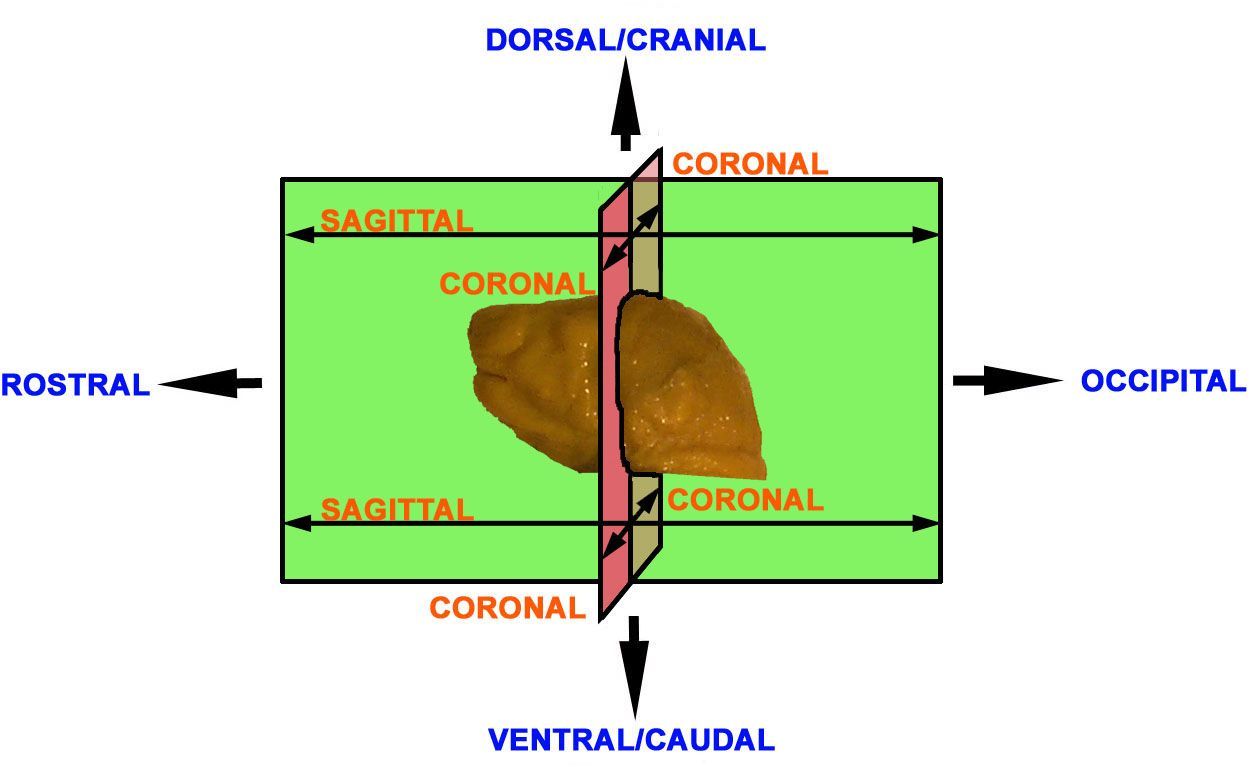
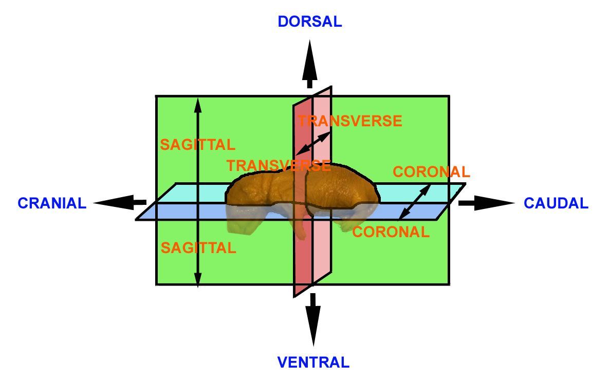
Anatomical planes
It is considered to be important that the examiner has an appreciation of the terms used to describe the angles at which the specimen is viewed and which can be used to accurately describe observations.
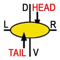
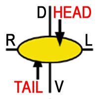
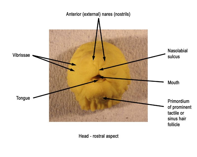
Anatomical description requires the use of a series of terms to denote angles, views, axes, planes and directions. As with the nomenclature of structures, there are various conventions. The terms cranial (or dorsal) and caudal (or ventral) are used to denote the upper and lower regions of the head, respectively. "Rostral" is used to denote the nasal extremity of the head and "occipital" to denote the back of the head (the region at the junction of the head and neck).
The rat head and body
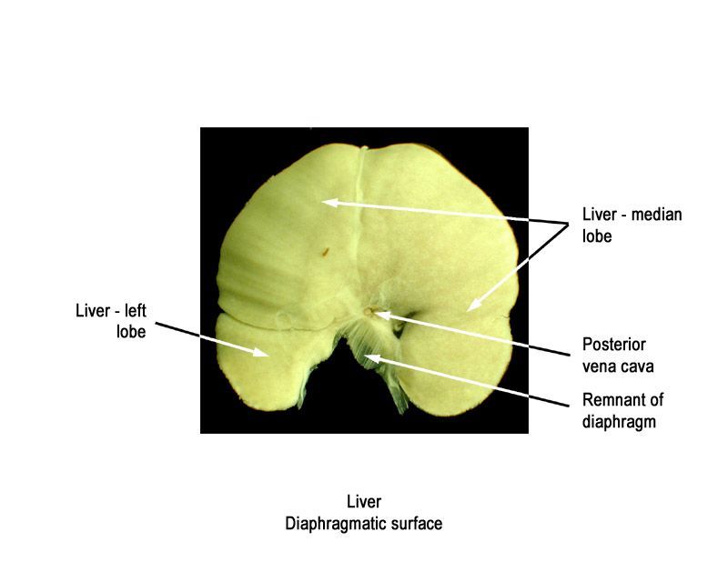
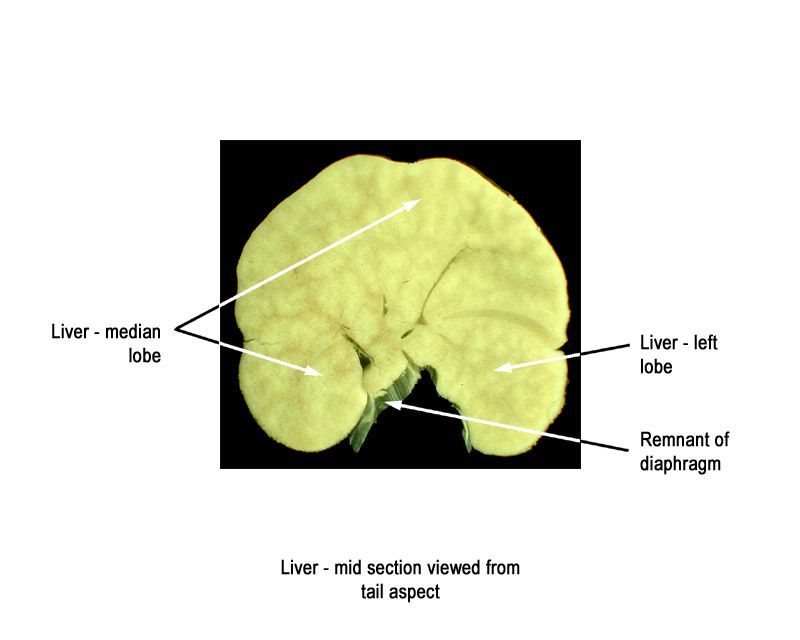
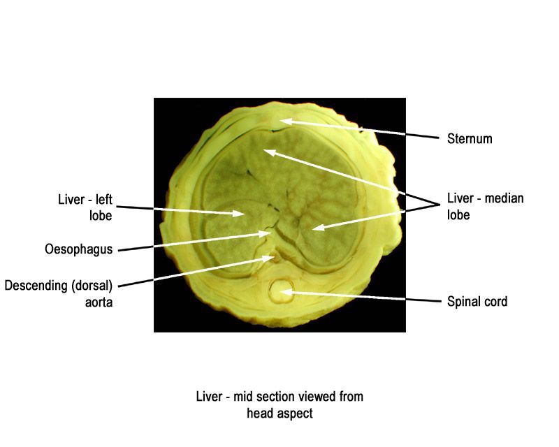
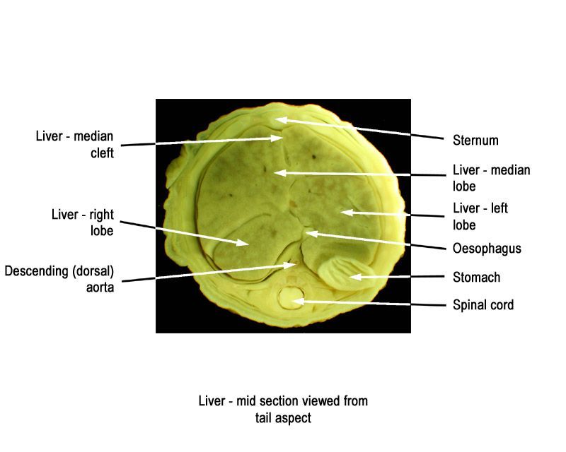
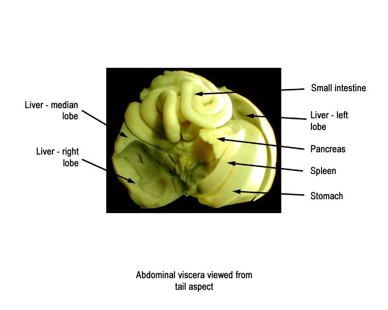
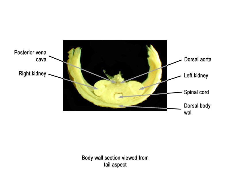
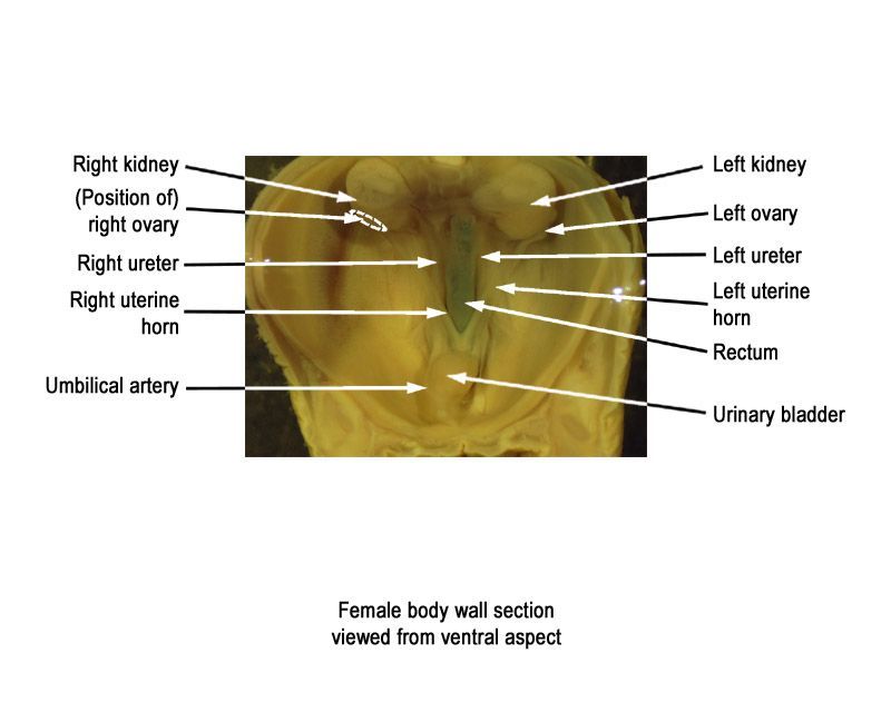
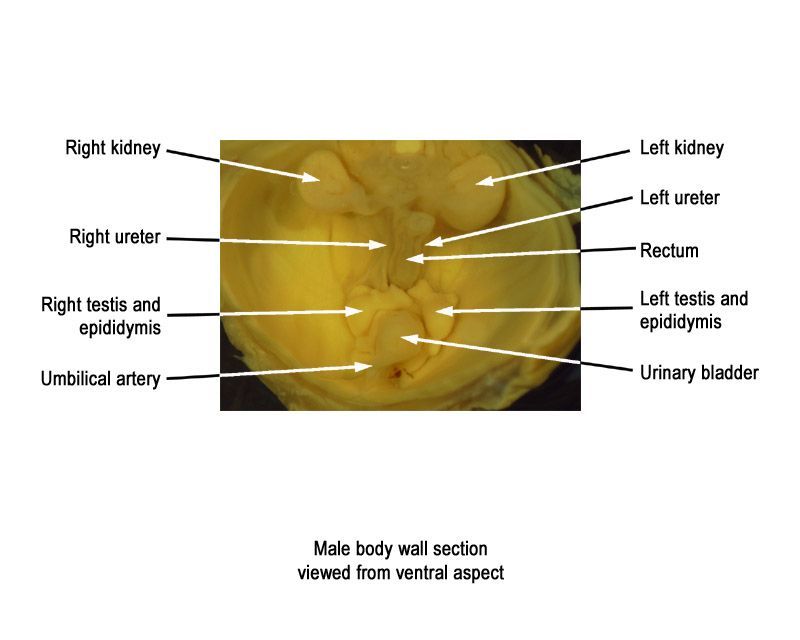
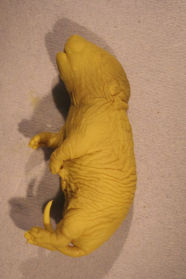
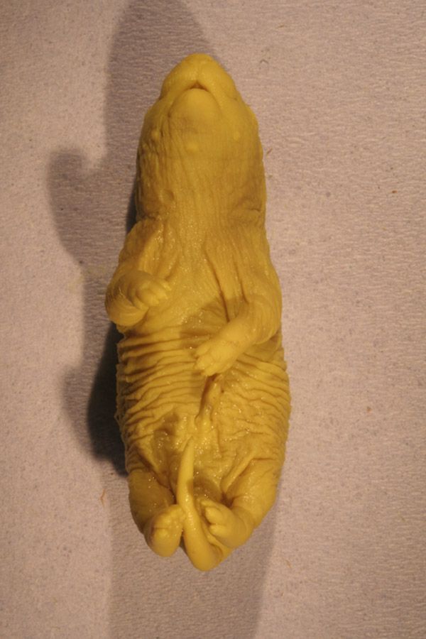
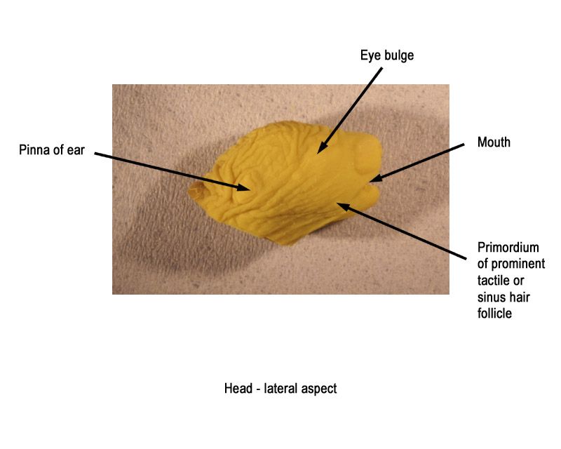
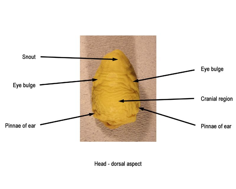
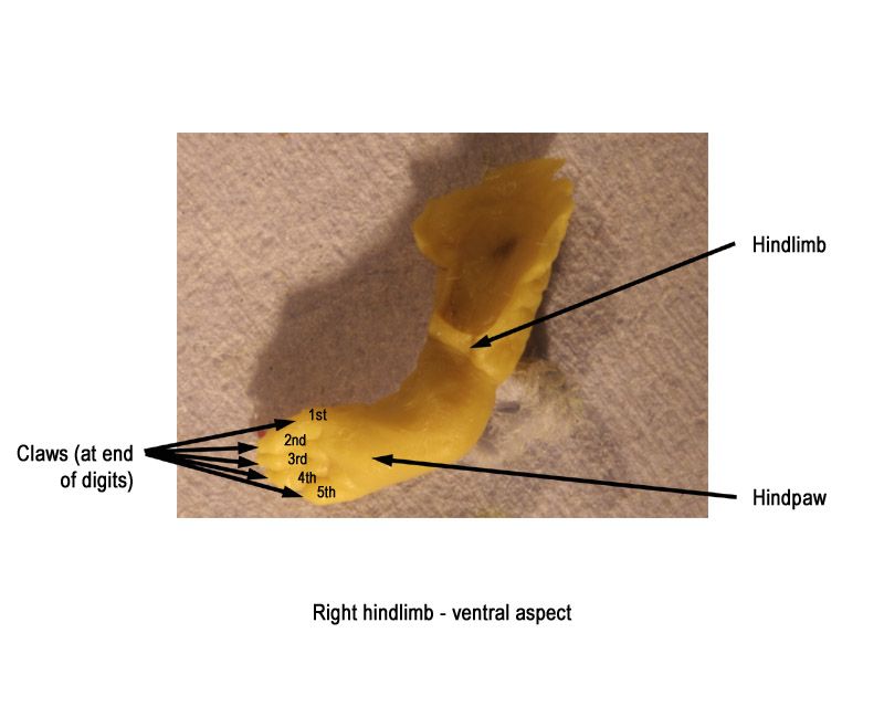
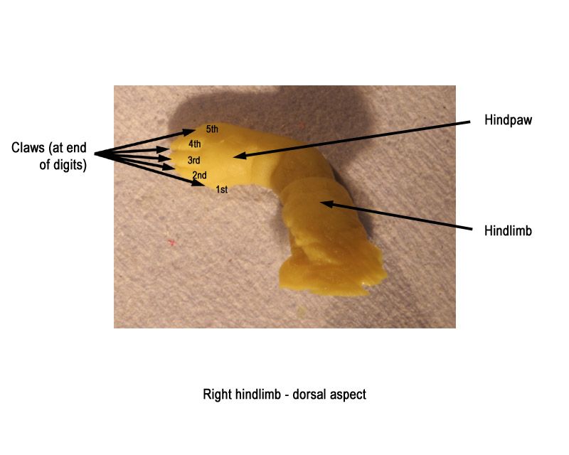
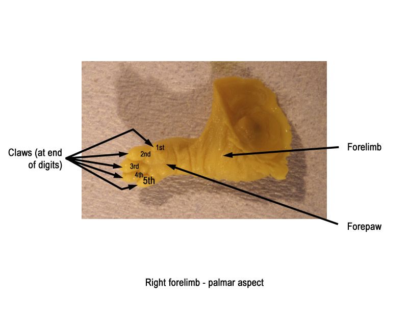
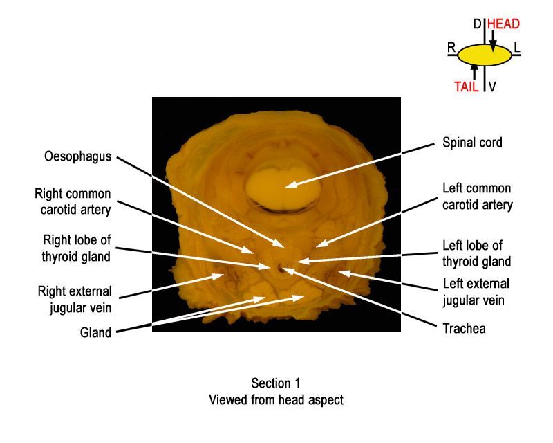
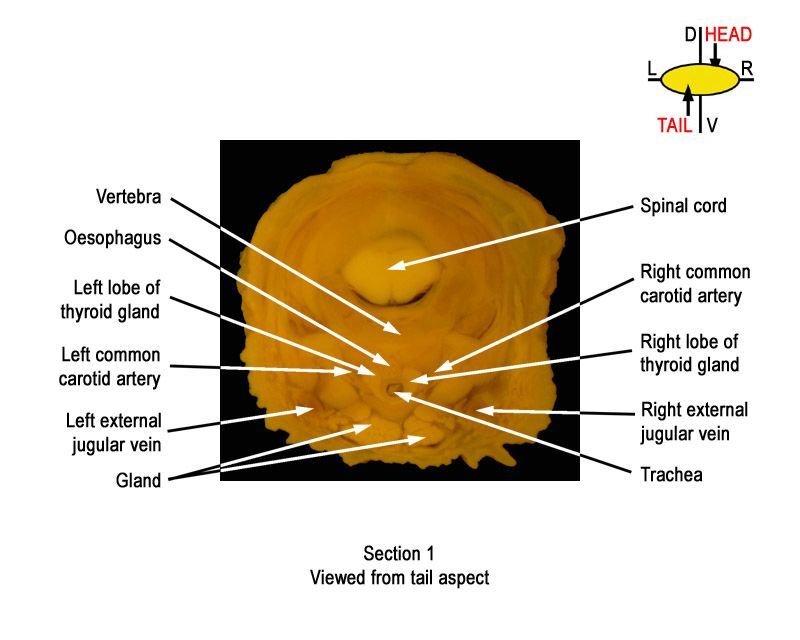
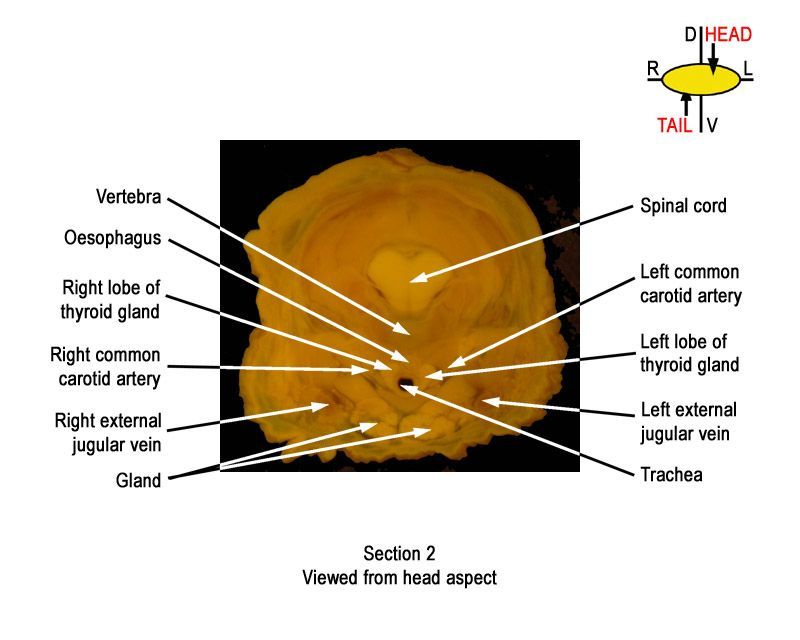
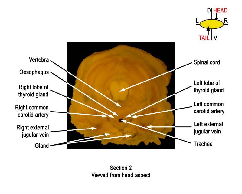
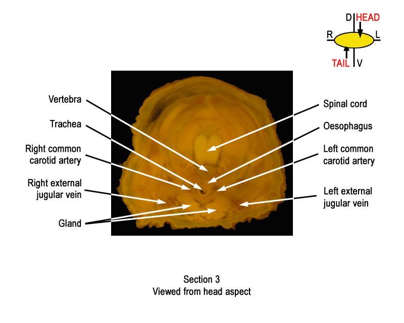
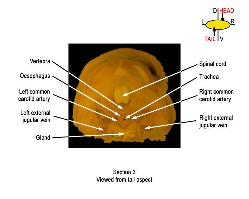
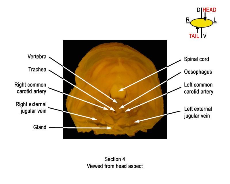
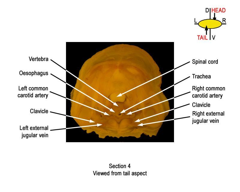
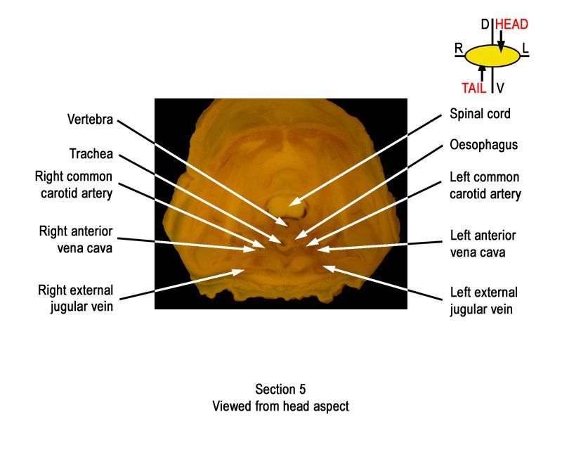
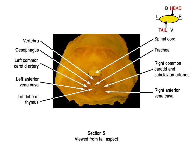
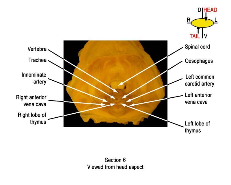
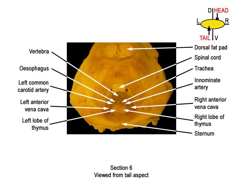
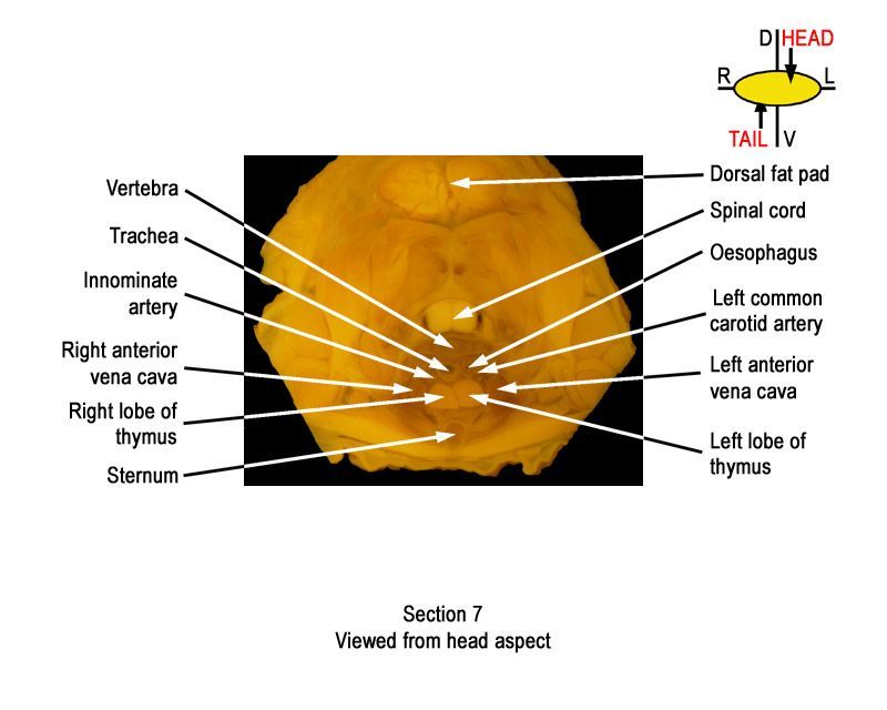
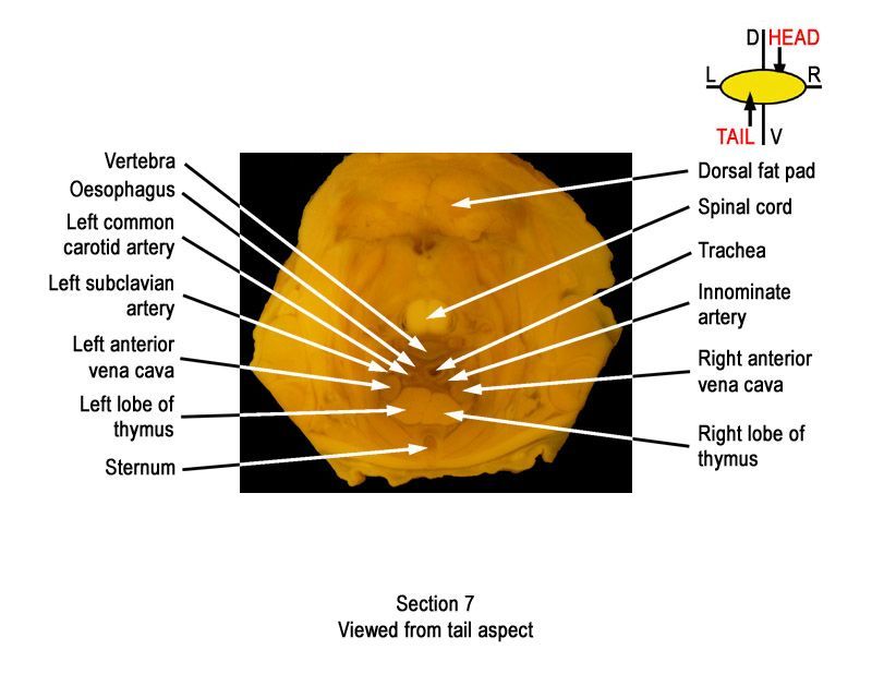
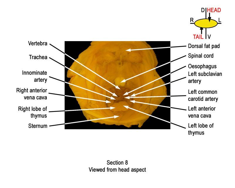
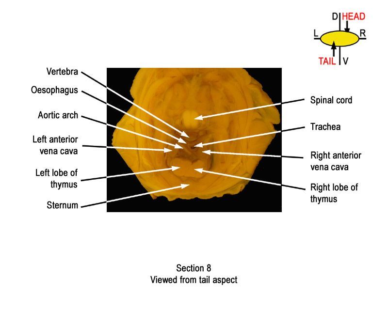
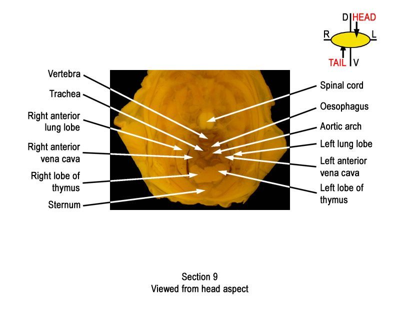
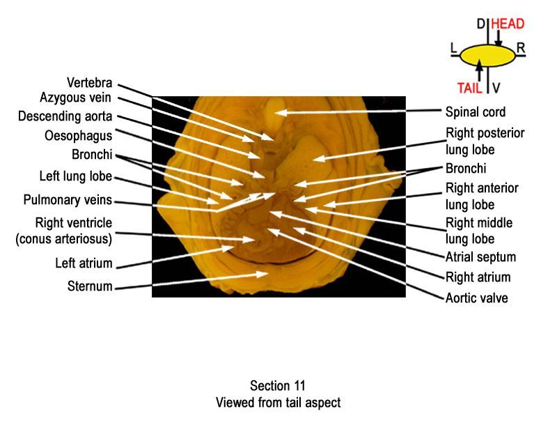
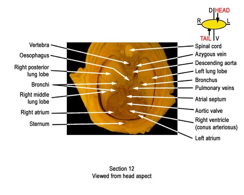
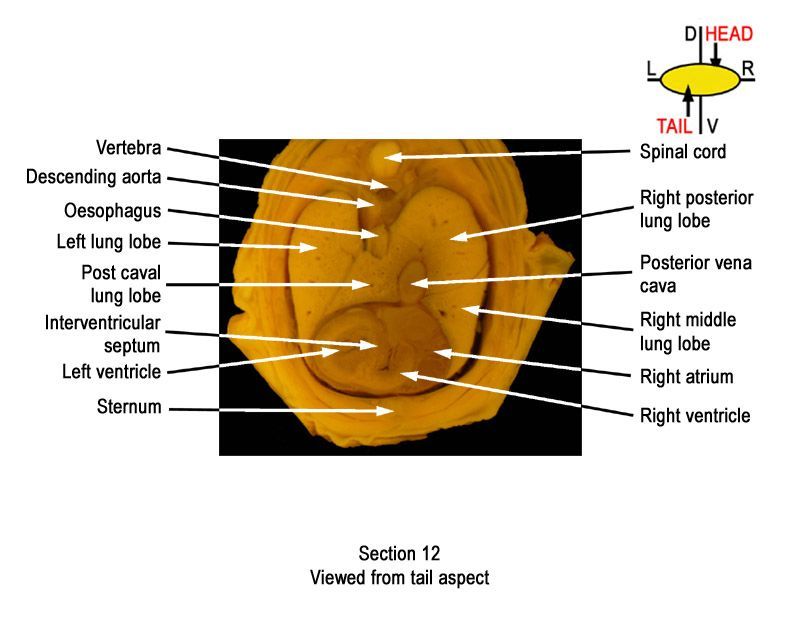
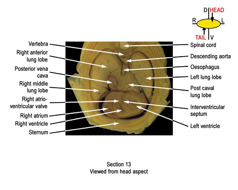
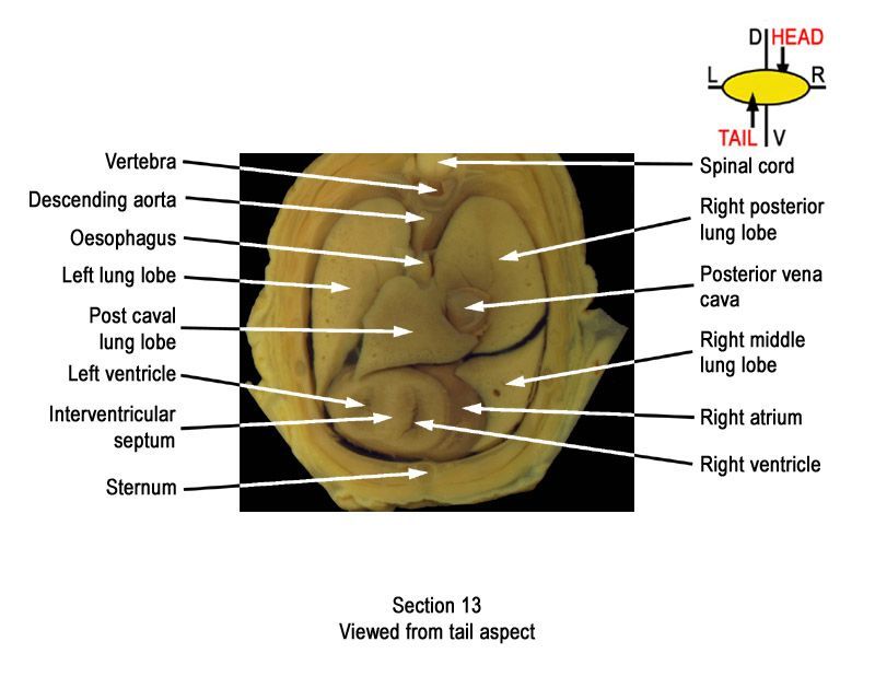
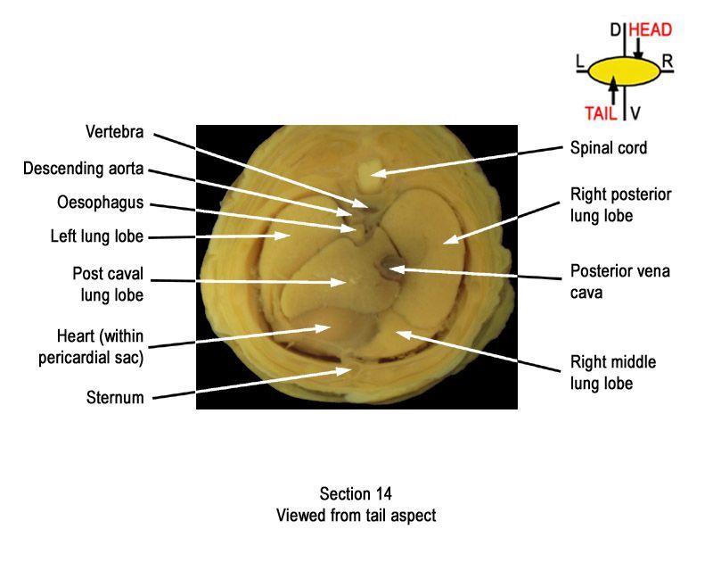
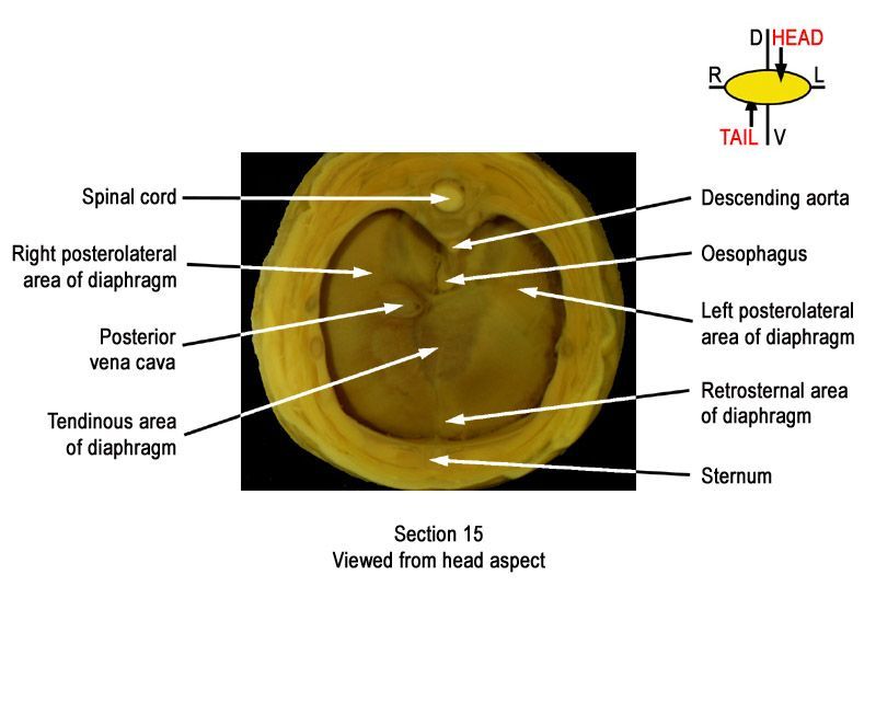
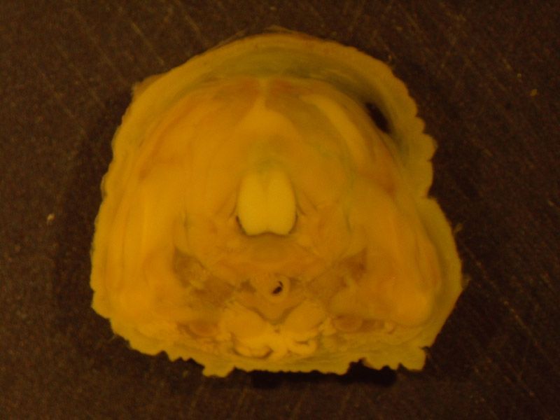
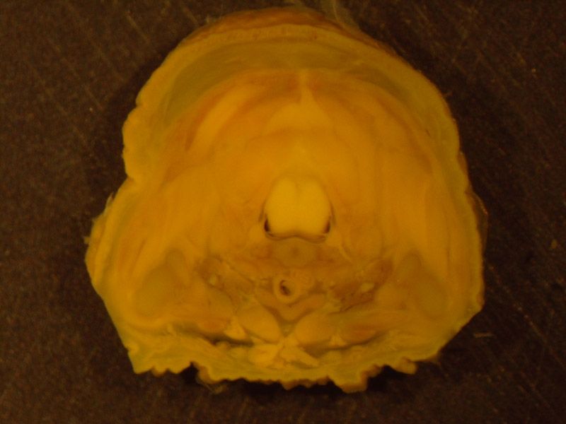
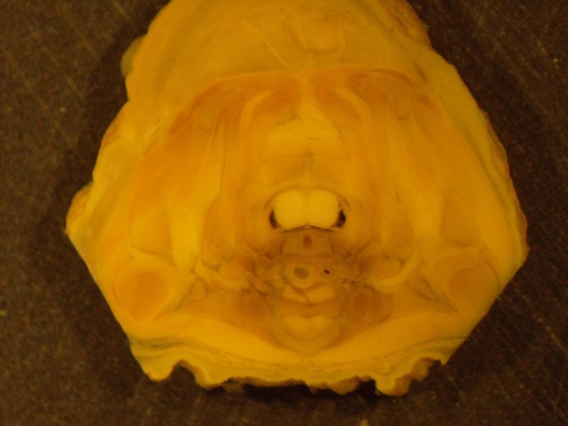
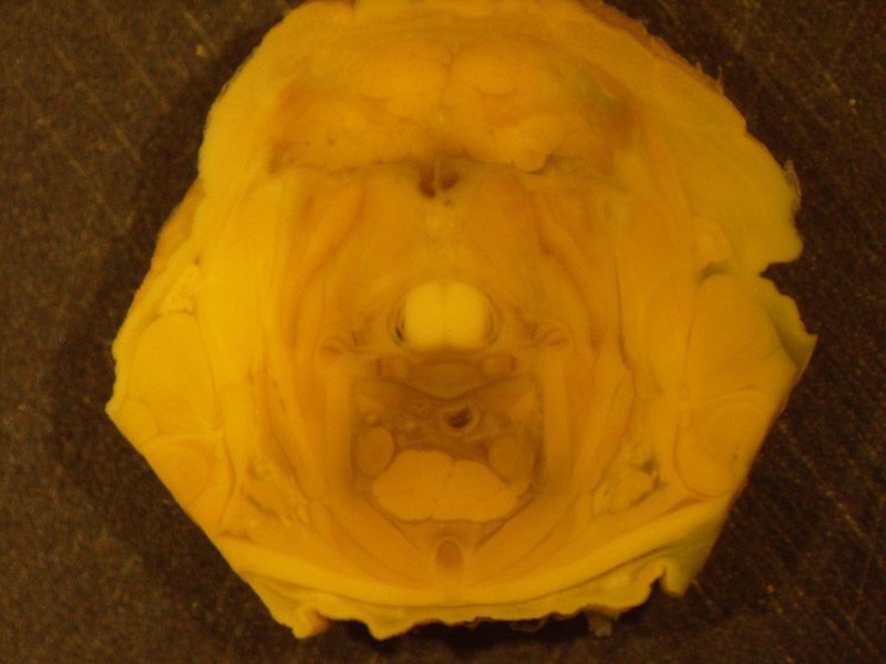
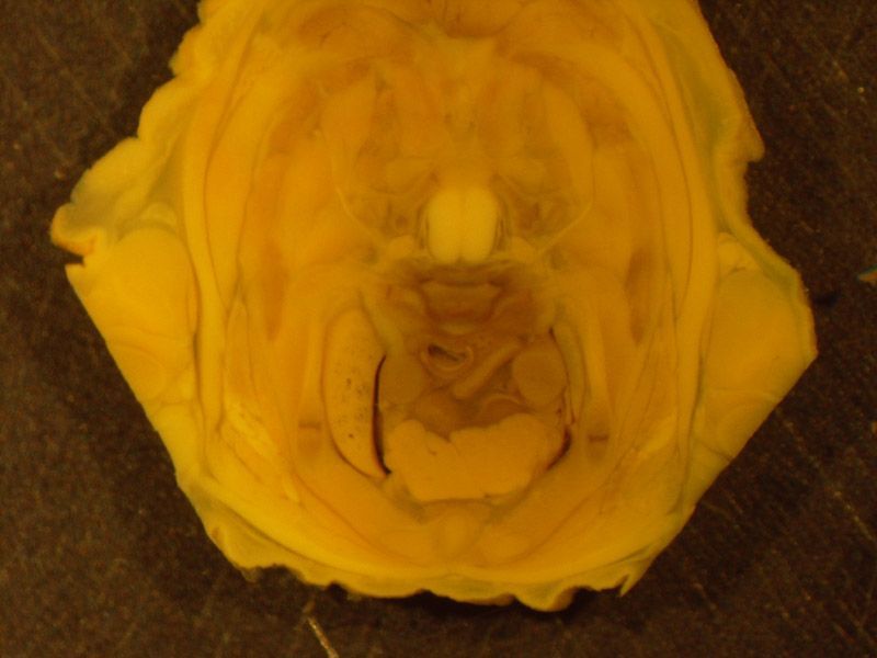
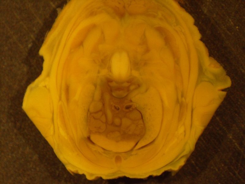
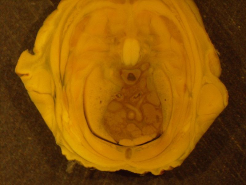
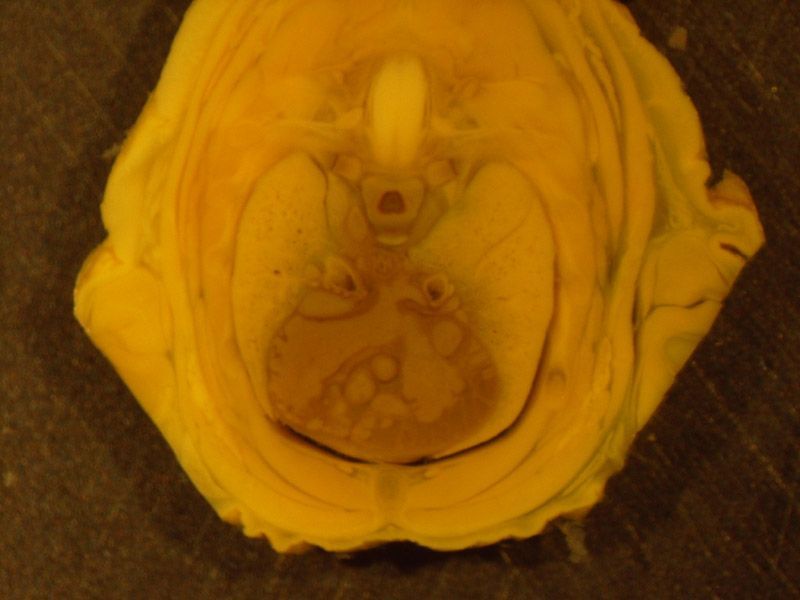
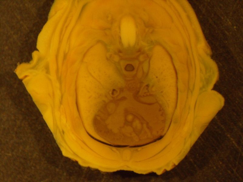
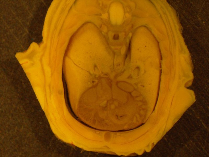
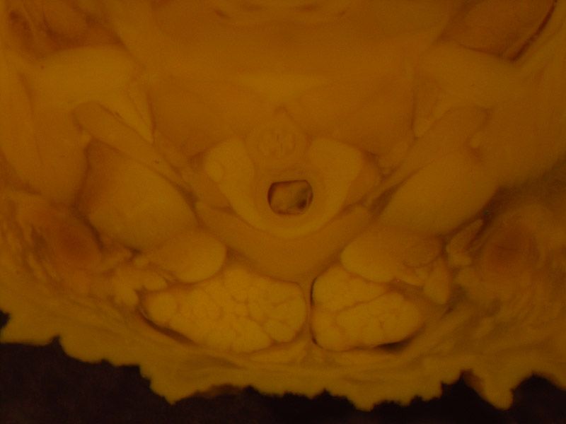
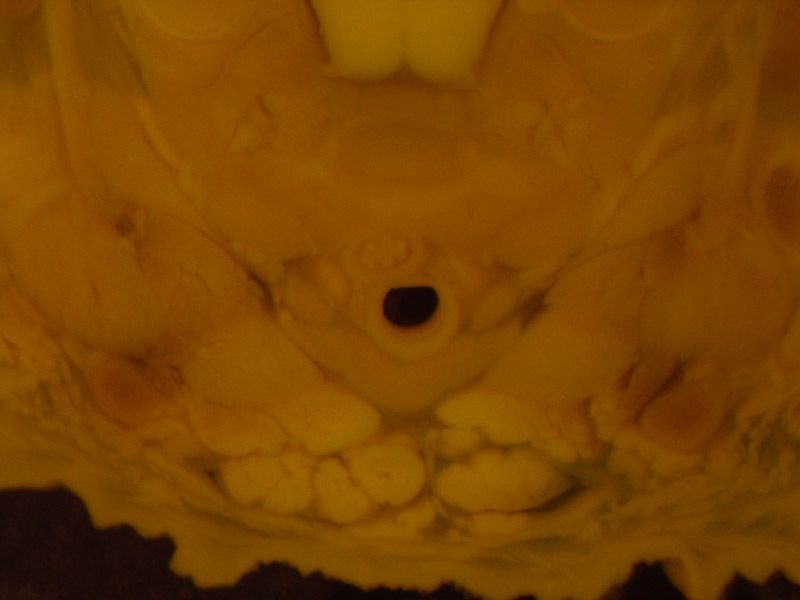
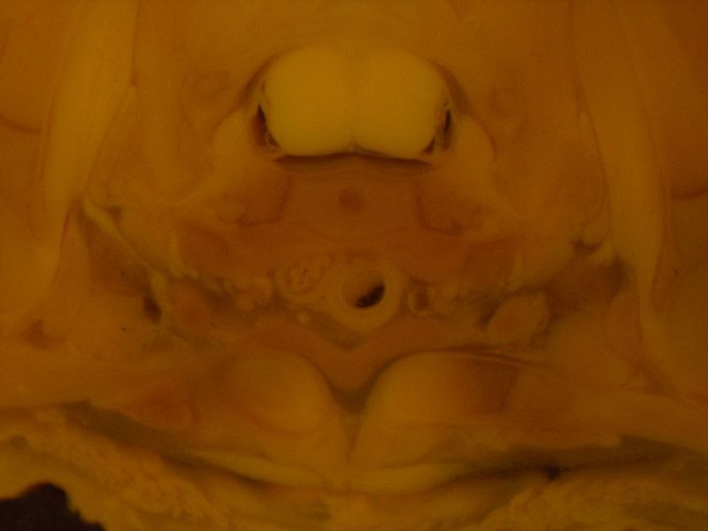
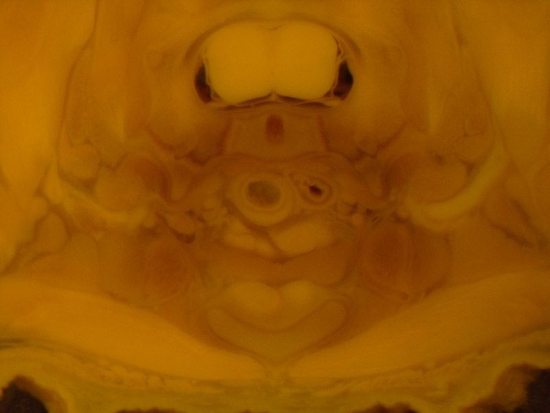
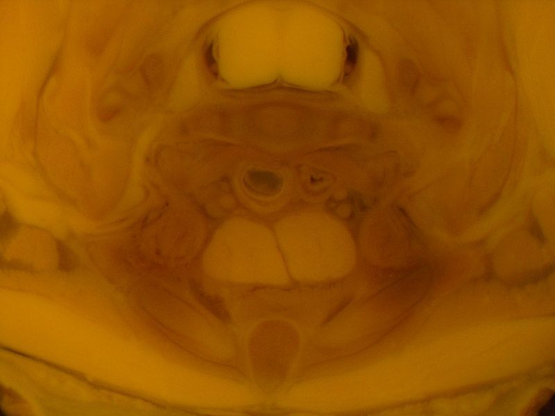
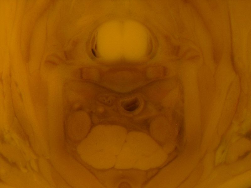
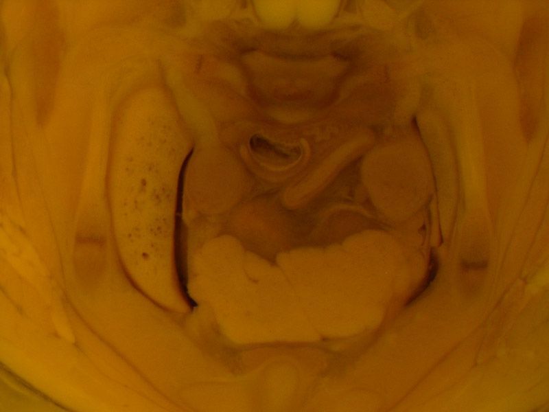
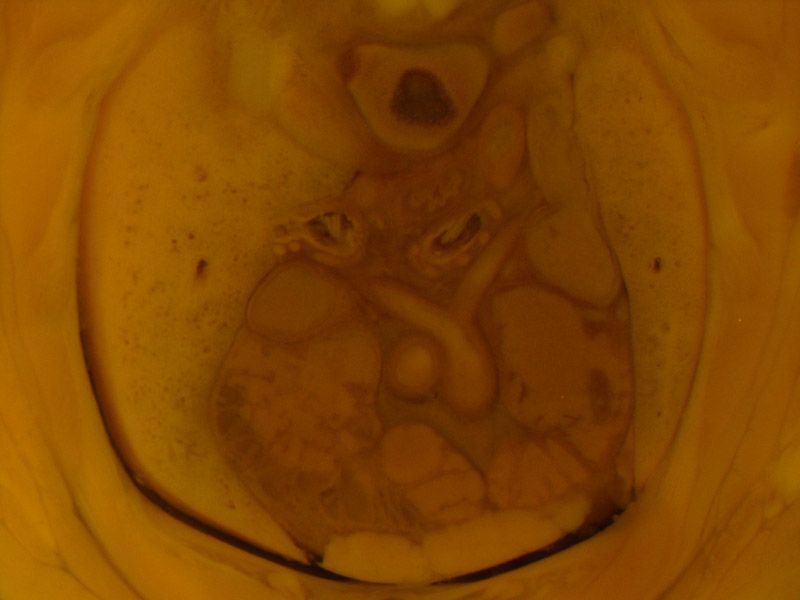
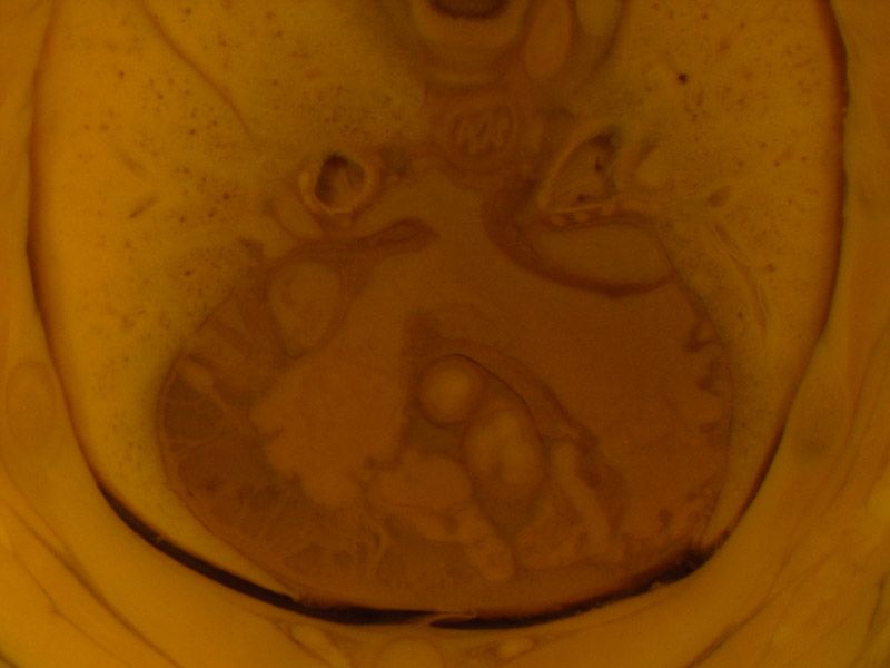
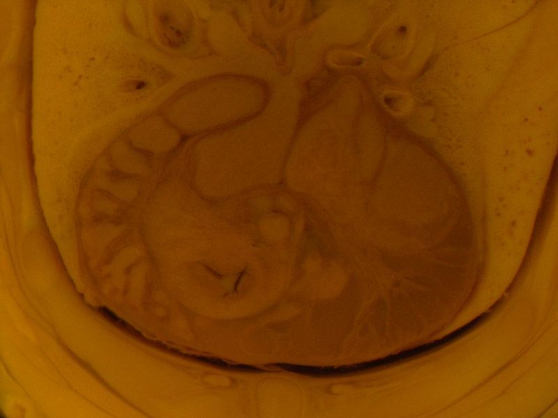
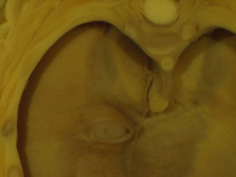
The images below show the normal appearance of Bouin's fluid fixed head and body Wilson sections in specimens at Day 21 of gestation (day mating observed = Day 0).
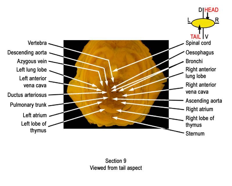
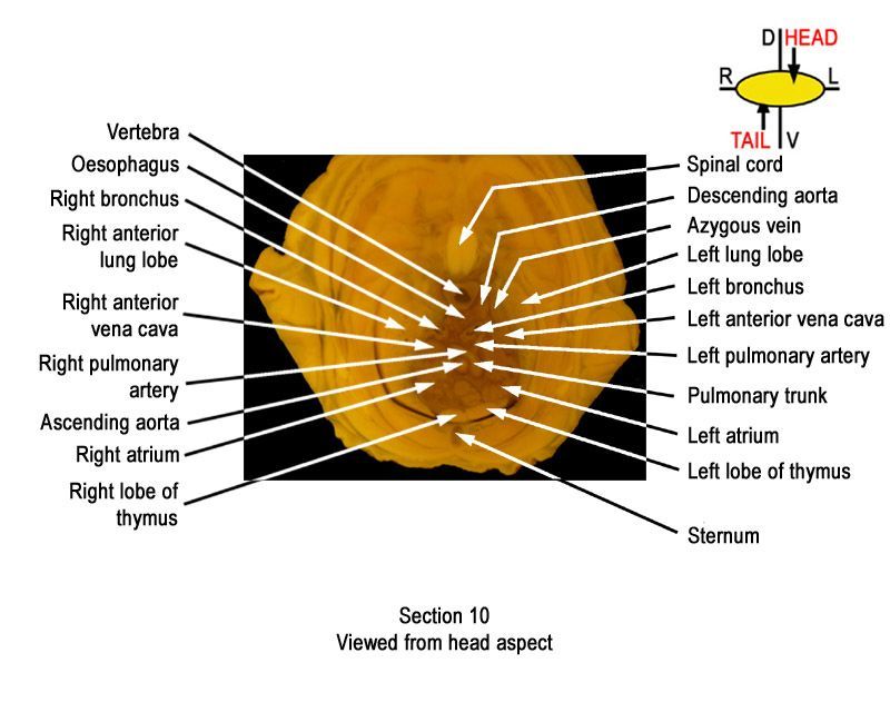
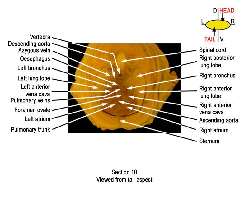
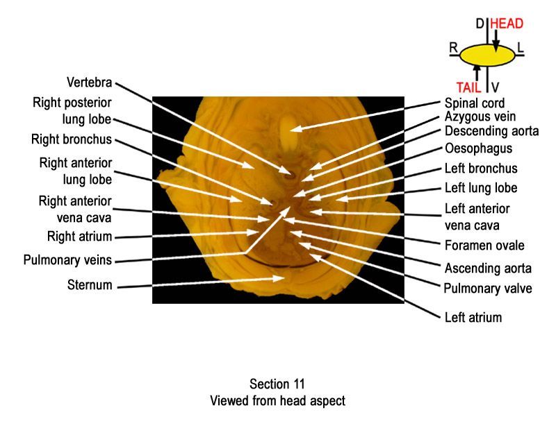
It is essential that both sides of each section is examined so that structures that exist is several sections can be visualised by the examiner in 3D.
It is recommended that all structures listed on the The International Register of Fetal Morphologists (IRFM) Expected Minimum Structure List for Fetal Morphology Examinations document be examined and that the Fixed head and body examination - key structures and notes document be used as a reference.
Learning objective: Compare the diagrams with your own specimens and identify all of the structures that have been labelled.
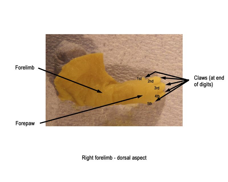
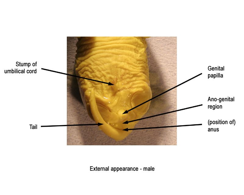
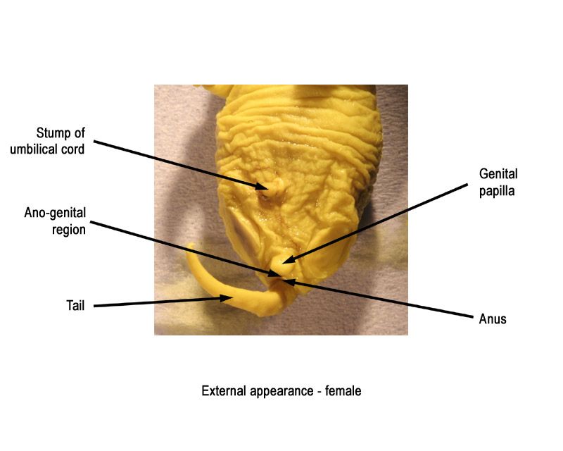
External appearance - click on an image below to open the gallery
Body (cervical/thoracic) sections - Labelled Images, Unlabelled Images and 'Close up' Images - click on an image to open the gallery


Body (abdominal) sections - click on an image to open the gallery