NORMAL APPEARANCE OF THE RABBIT SKELETON
Anatomical planes
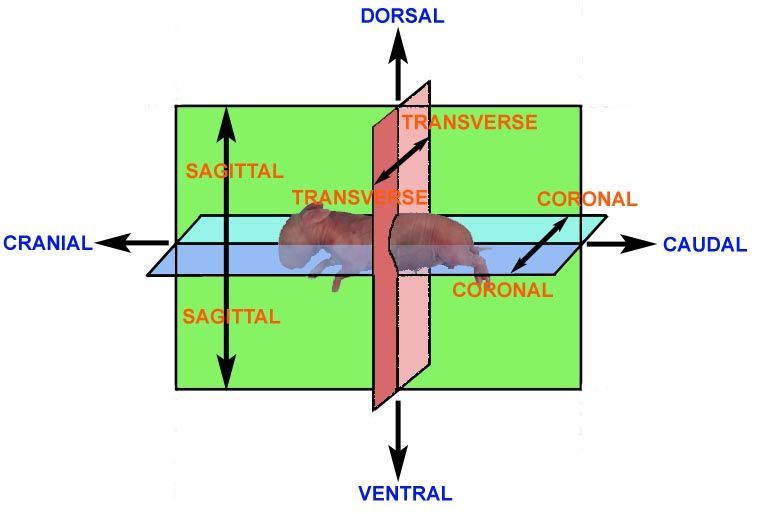
It is considered to be important that the examiner has an appreciation of the terms used to describe the angles at which the specimen is viewed and which can be used to accurately describe observations.
Anatomical description requires the use of a series of terms to denote angles, views, axes, planes and directions. As with the nomenclature of the bones, there are various conventions. The terms cranial and caudal are used to denote the upper (head) and lower (tail) body regions, respectively; dorsal and ventral are used to denote the front and back aspects of the body. "Rostral" is used to denote the nasal extremity of the head and "occipital" to denote the back of the head (the region at the junction of the head and neck).
THE RABBIT SKELETON
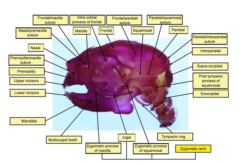
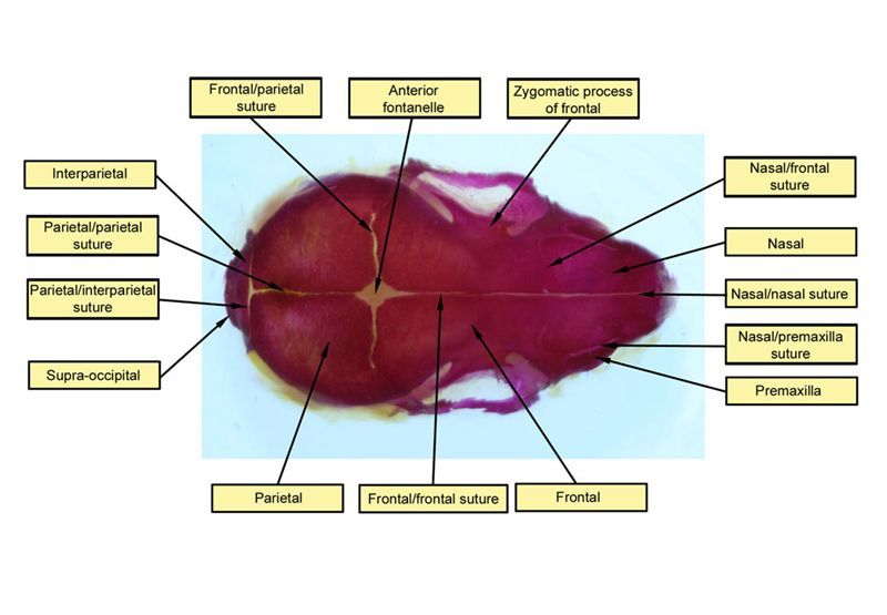
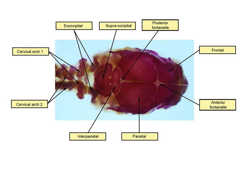
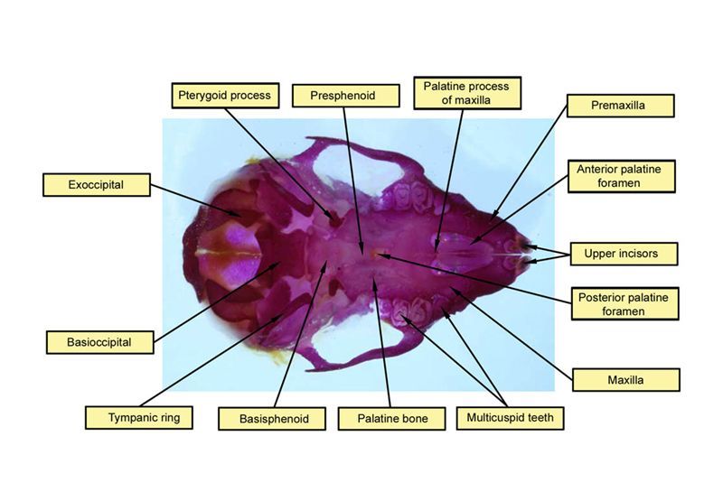
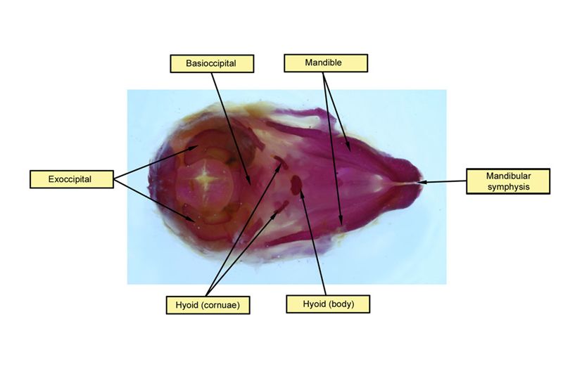
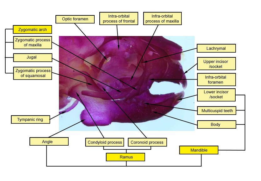
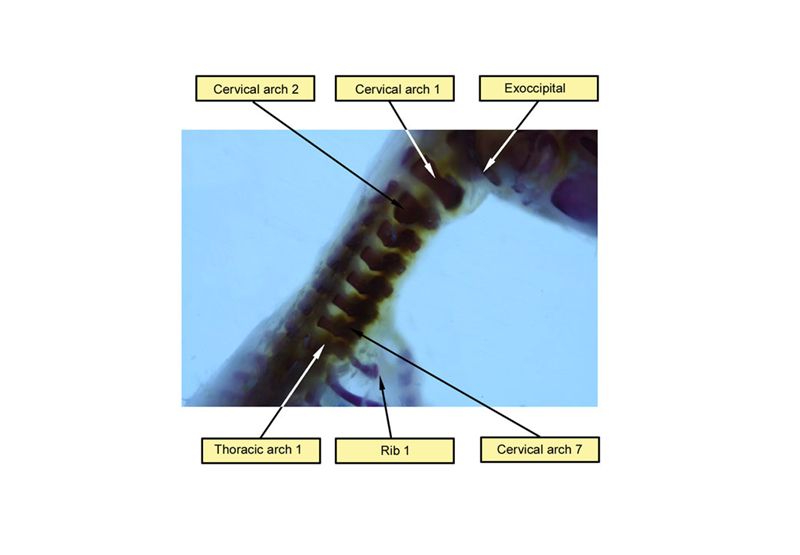
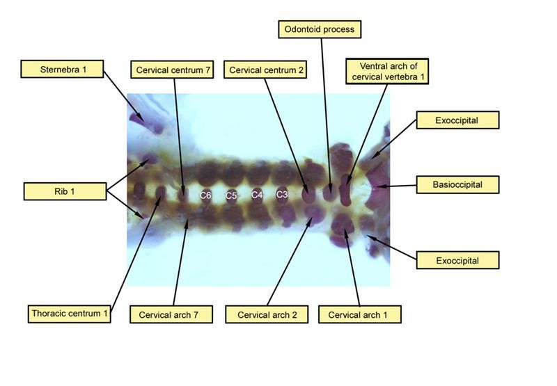
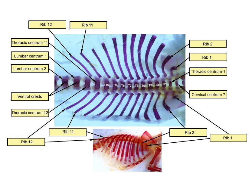
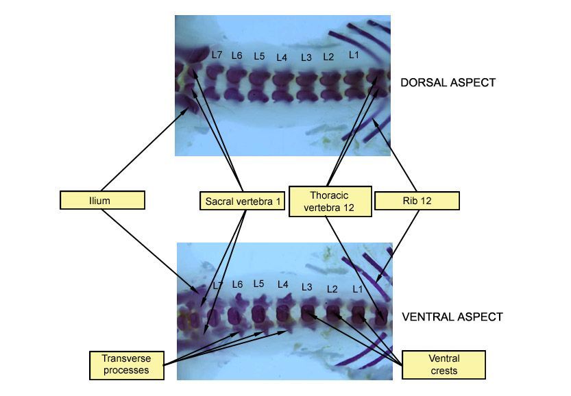
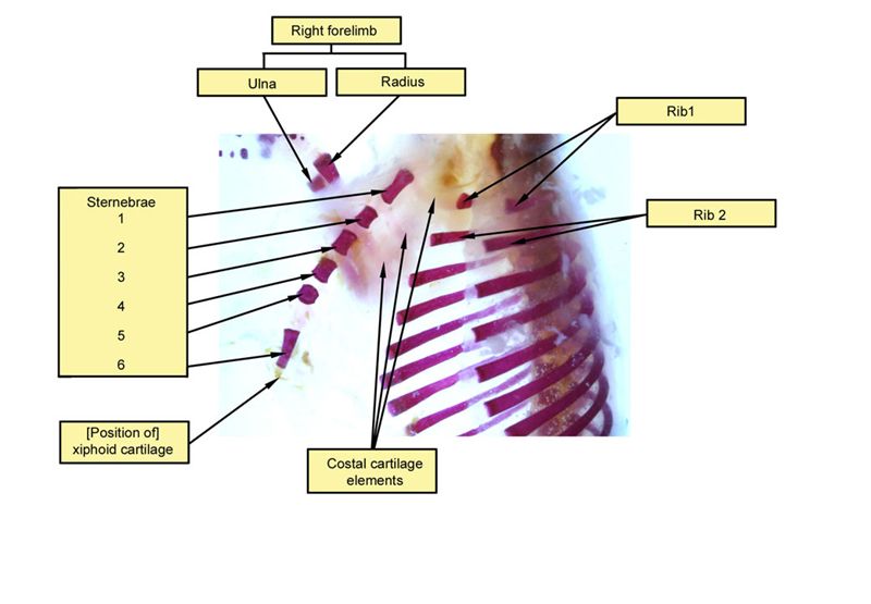
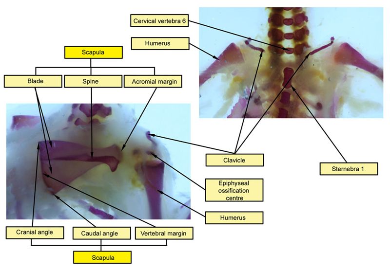
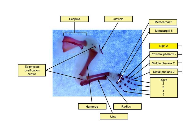
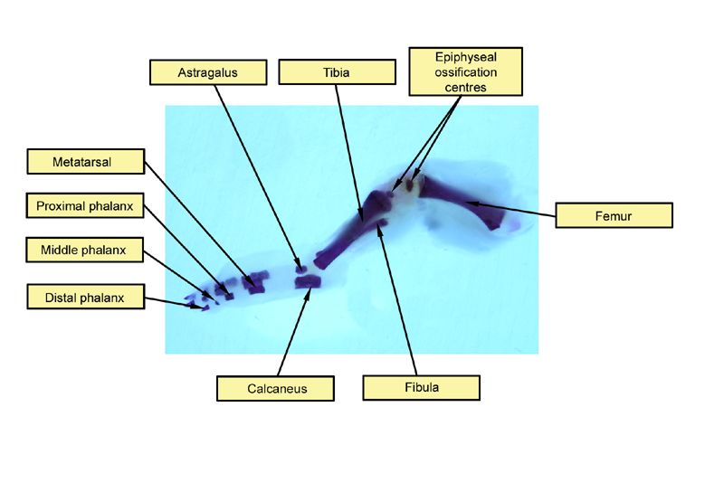
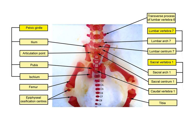
The images below show the normal appearance of skeletal elements in specimens at Day 29 of gestation (day mating observed = Day 0).
It is essential that the skeleton is viewed from all angles, both to minimize the possibility of optical illusions and to maximize the opportunity for detection of any unusual features. While it is also recommended that certain features be finally evaluated consistently from one view (e.g. the vertebral arches could be evaluated primarily from the dorsal view), this must not preclude examination also from other angles.
It is recommended that all structures listed on the The International Register of Fetal Morphologists (IRFM) Expected Minimum Structure List for Fetal Morphology Examinat document be examined and that the Skeletal examination - key structures and notes document be used as a reference.
Learning objective: Compare the diagrams with your own specimens and identify all of the structures that have been labelled.
Click on an image below to open the photo gallery