NORMAL APPEARANCE OF THE RAT SKELETON
Anatomical planes
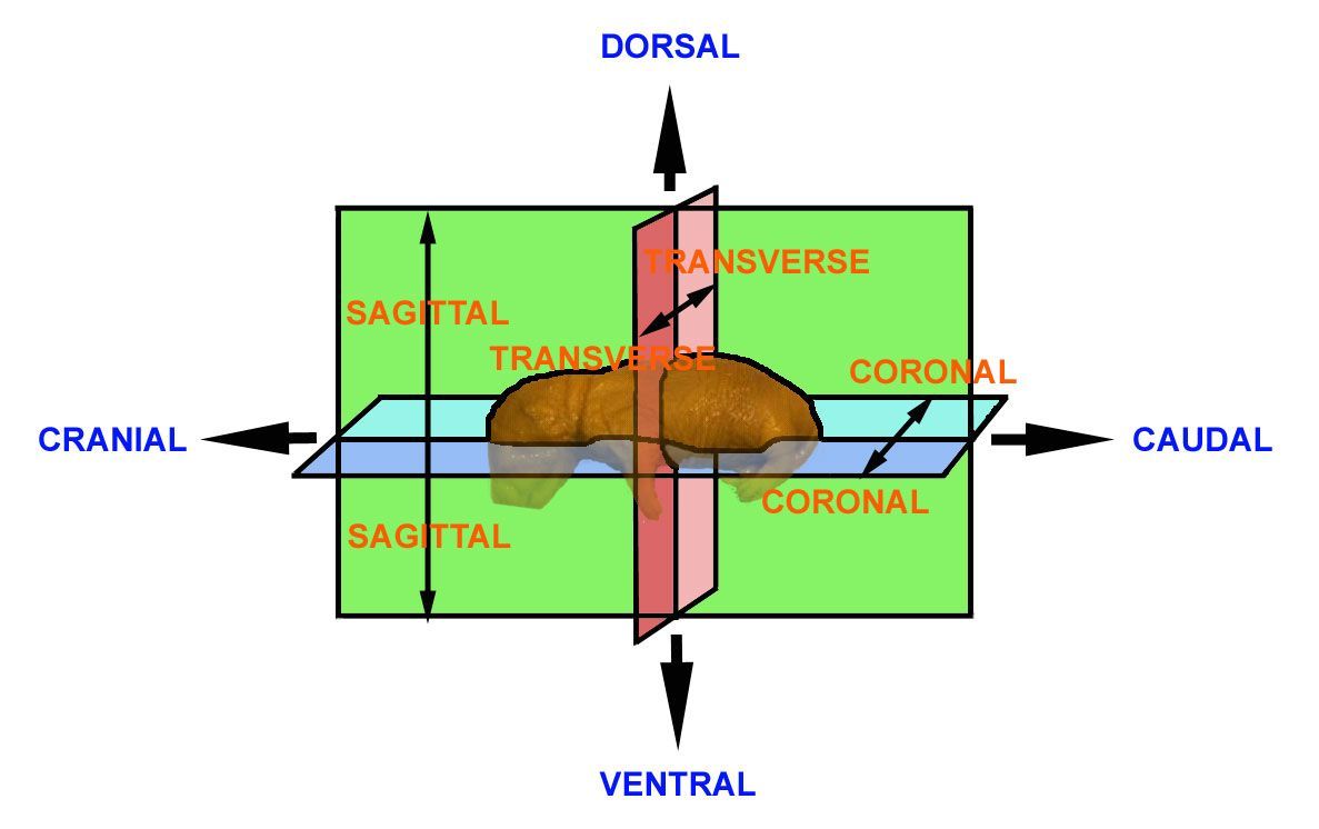
It is considered to be important that the examiner has an appreciation of the terms used to describe the angles at which the specimen is viewed and which can be used to accurately describe observations.
Anatomical description requires the use of a series of terms to denote angles, views, axes, planes and directions. As with the nomenclature of the bones, there are various conventions. The terms cranial and caudal are used to denote the upper (head) and lower (tail) body regions, respectively; dorsal and ventral are used to denote the front and back aspects of the body. "Rostral" is used to denote the nasal extremity of the head and "occipital" to denote the back of the head (the region at the junction of the head and neck).
The rat skeleton
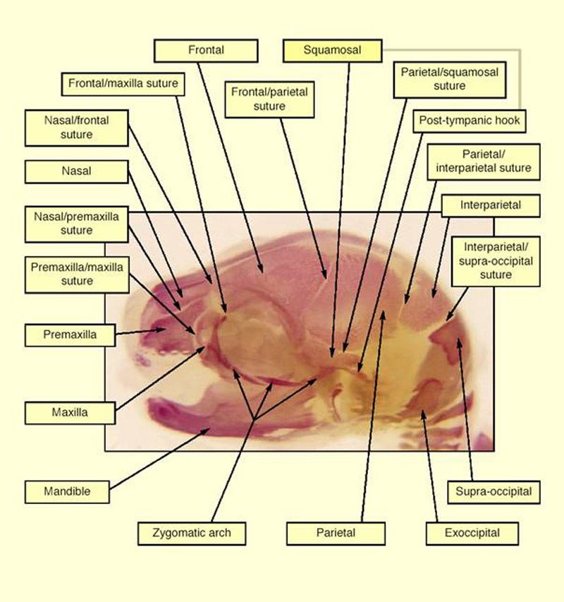
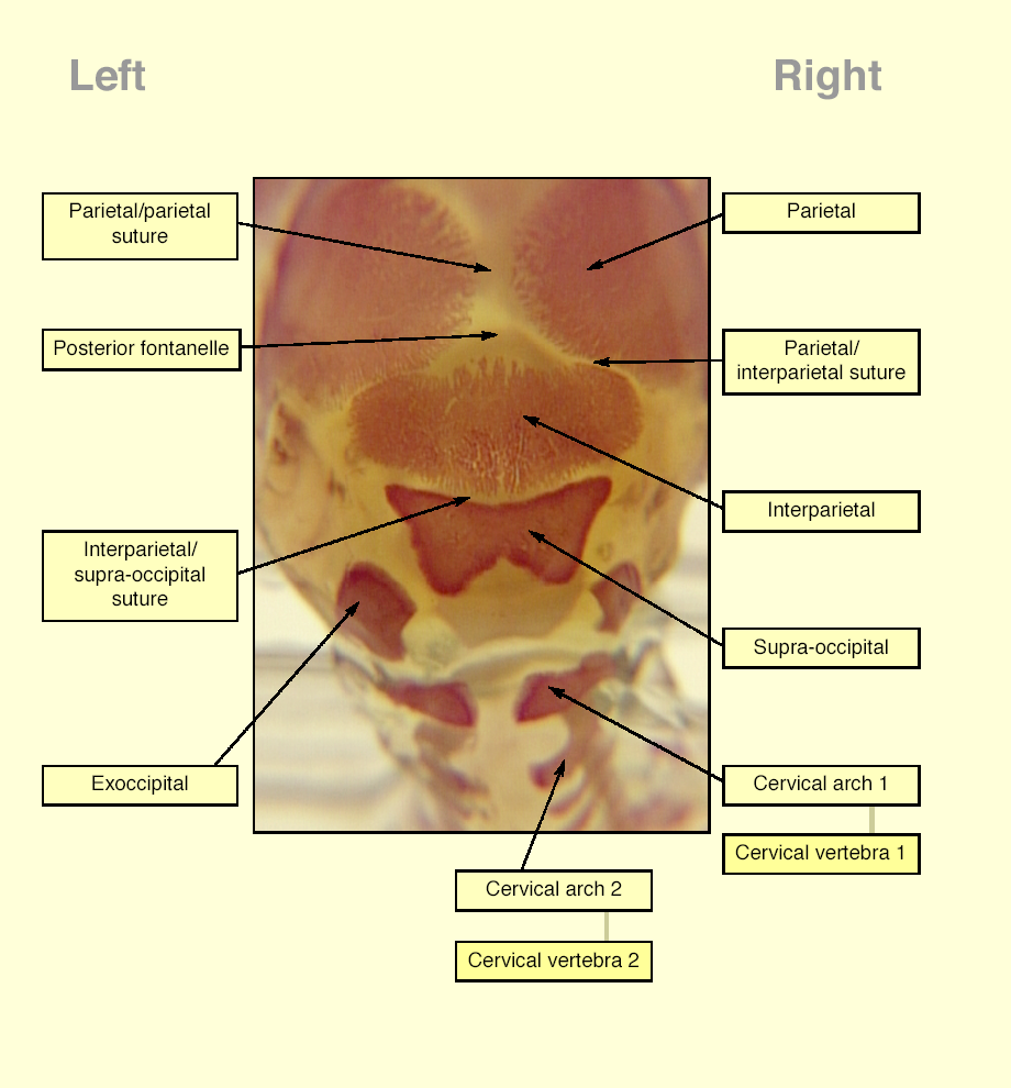
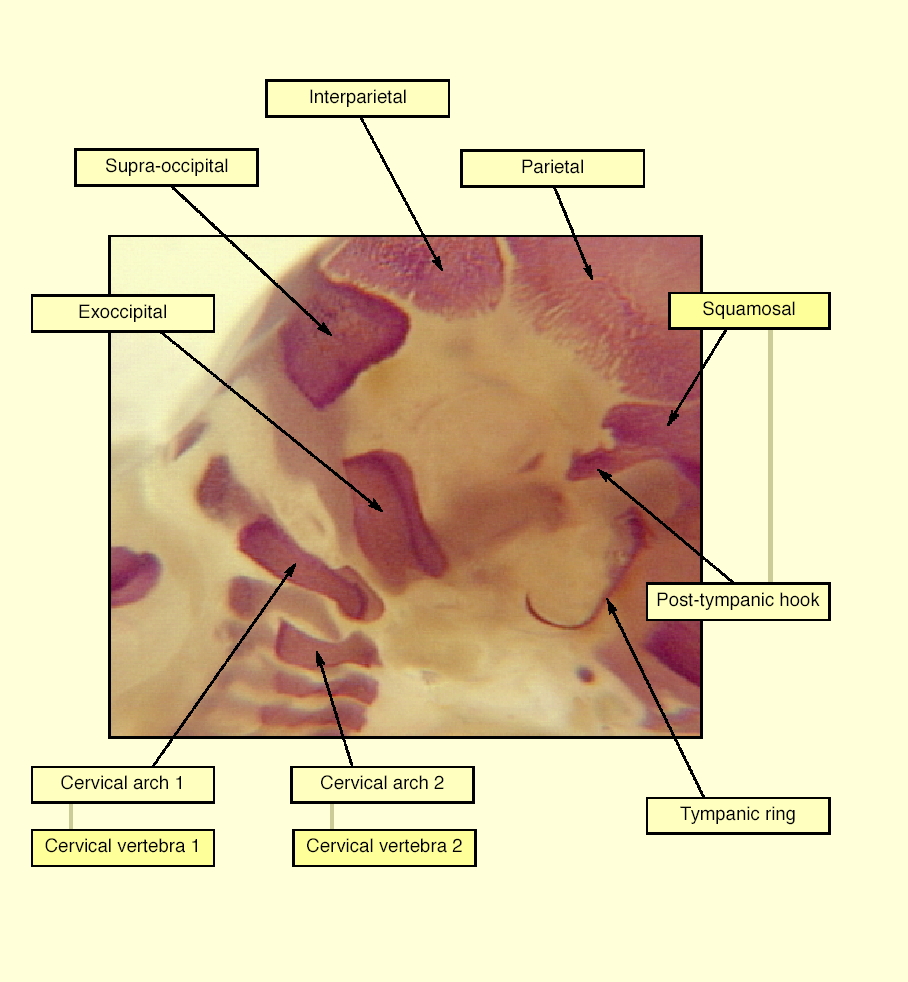
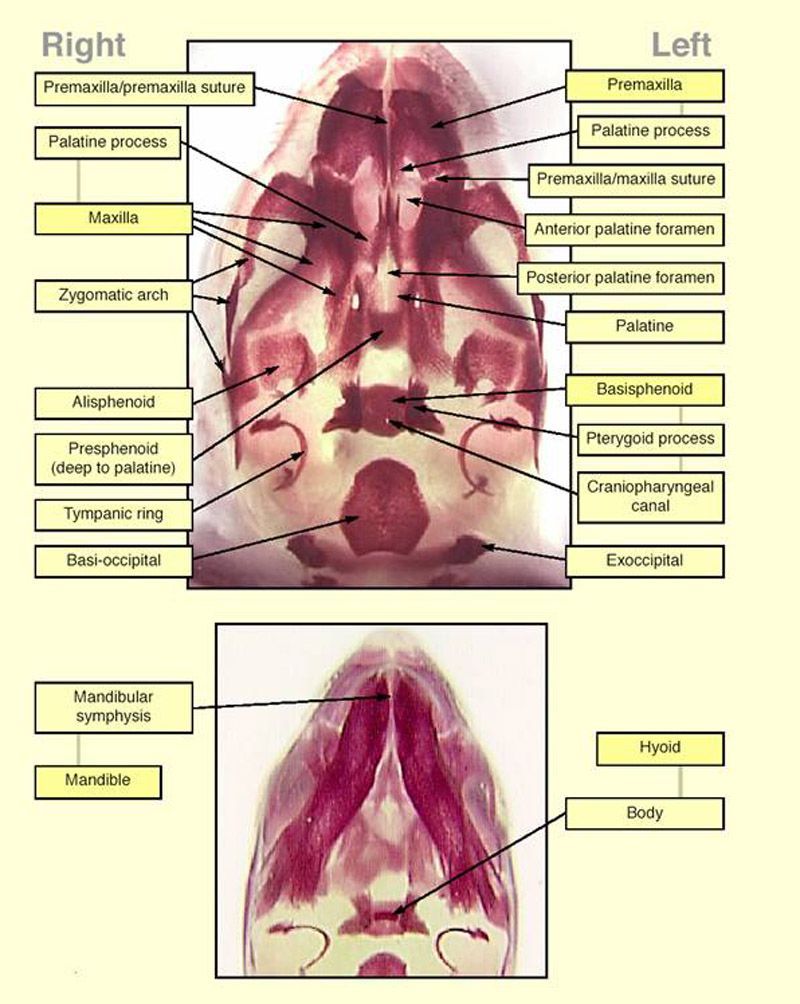
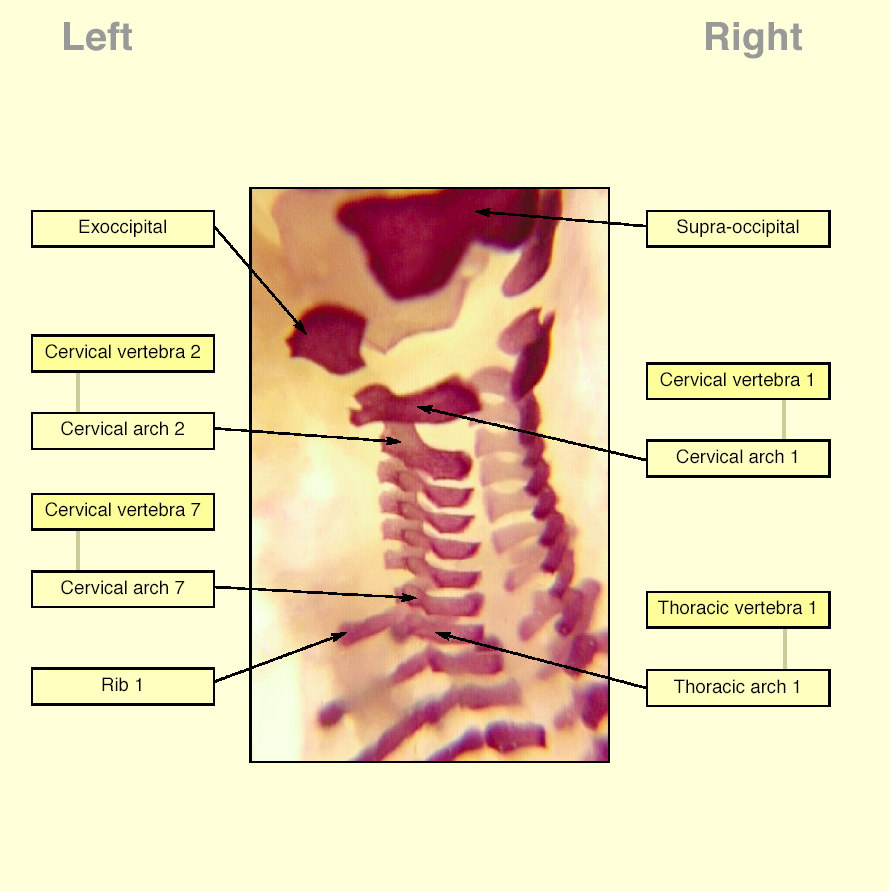
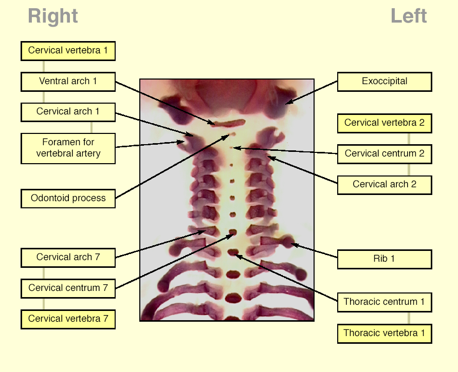
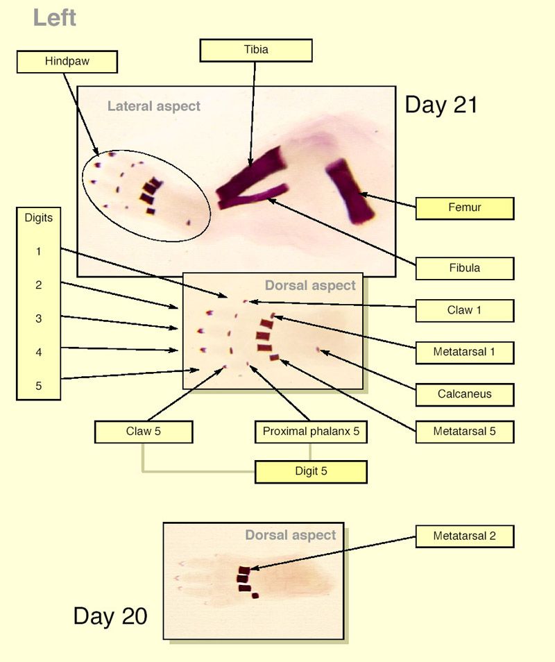
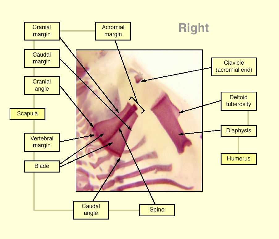
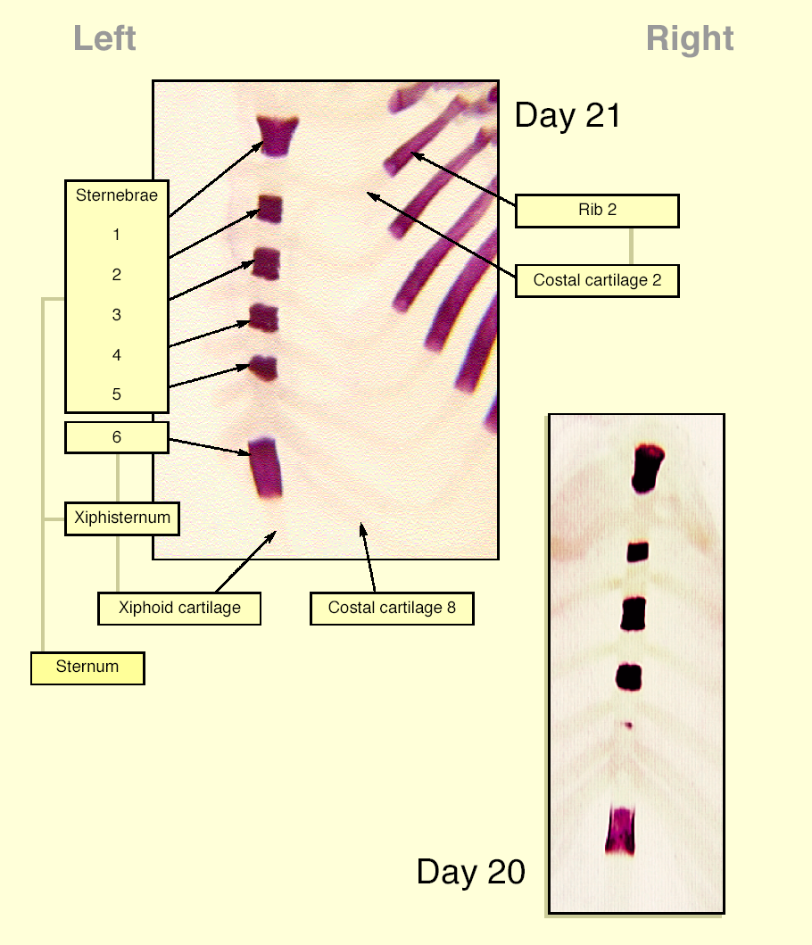
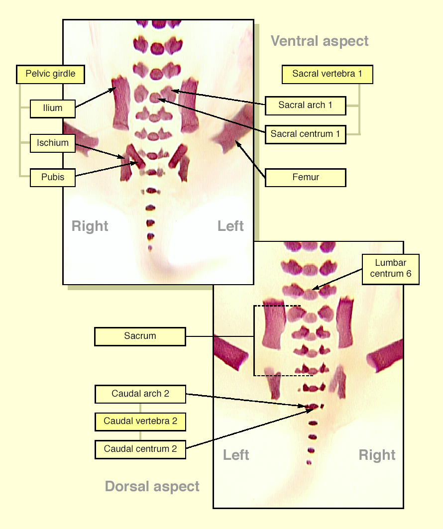
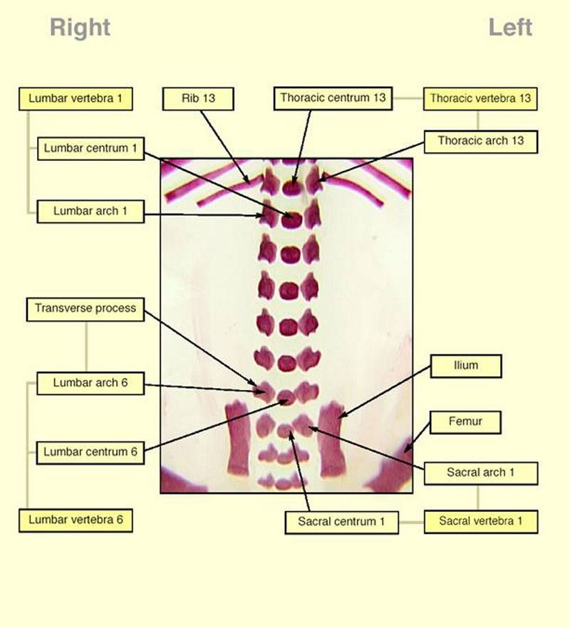
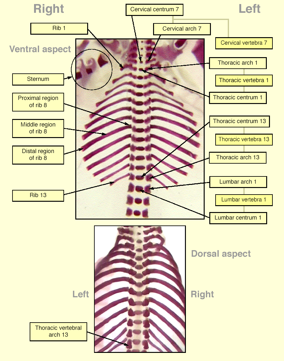
The images below show the normal appearance of skeletal elements in specimens at Day 20 and 21 of gestation (day evidence of mating observed = Day 0).
It is essential that the skeleton is viewed from all angles, both to minimize the possibility of optical illusions and to maximize the opportunity for detection of any unusual features. While it is also recommended that certain features be finally evaluated consistently from one view (e.g. the vertebral arches could be evaluated primarily from the dorsal view), this must not preclude examination also from other angles.
It is recommended that all structures listed on the The International Register of Fetal Morphologists (IRFM) Expected Minimum Structure List for Fetal Morphology Examinations document be examined and that the Skeletal examination - key structures and notes document be used as a reference.
Learning objective: Compare the diagrams with your own specimens and identify all of the structures that have been labelled.
An example in which calcium staining can result in the incorrect identification of structures relates to the claws. The claws are a derivative of the integument and have an outer layer of keratin, which takes up alizarin red S. The presence of stain at the distal extremity of a digit can be mistaken for ossification and thus can be assumed to represent the distal margin of the distal phalanx. Staining of the claws is characterized by a diffuse, crescent-shaped pattern at the distal margin of the claw; this is in contrast to the uniformly stained and cuboidal appearance of the phalangeal ossification centres lying towards the centre of the phalangeal cartilages.
Click on the images below to open the photo gallery