NORMAL APPEARANCE OF THE FRESH RABBIT FETUS
Anatomical planes
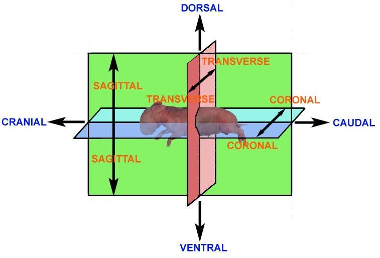
It is considered to be important that the examiner has an appreciation of the terms used to describe the angles at which the specimen is viewed and which can be used to accurately describe observations.
Anatomical description requires the use of a series of terms to denote angles, views, axes, planes and directions. As with the nomenclature of structures, there are various conventions. The terms cranial (or dorsal) and caudal (or ventral) are used to denote the upper and lower regions of the head, respectively. "Rostral" is used to denote the nasal extremity of the head and "occipital" to denote the back of the head (the region at the junction of the head and neck).
The rabbit fetus
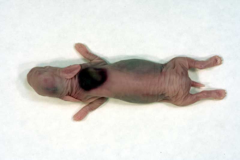
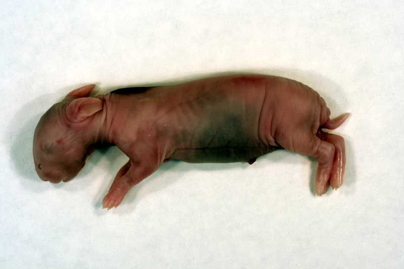
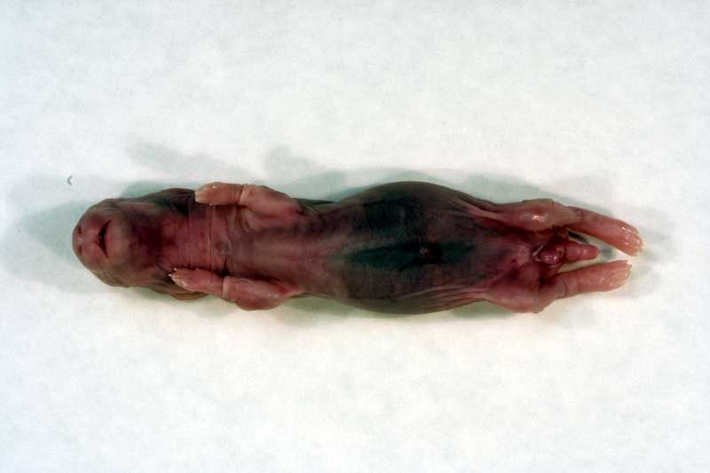
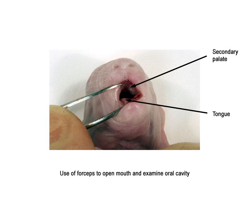
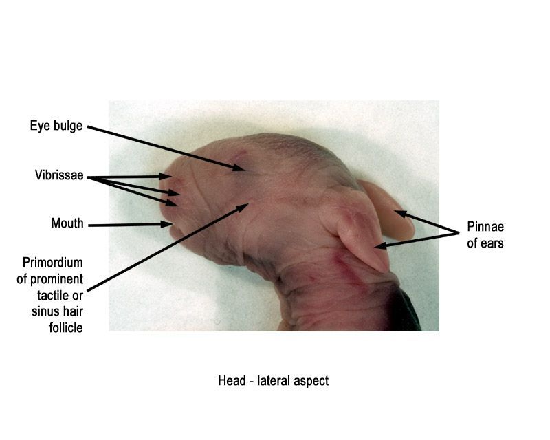
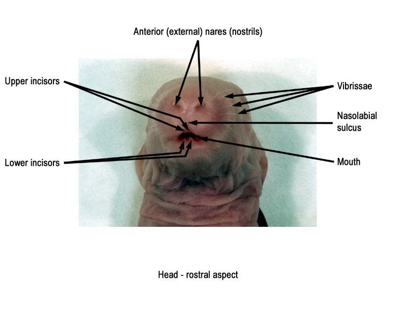
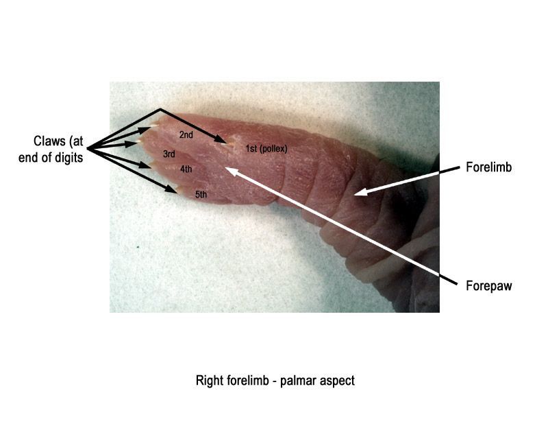
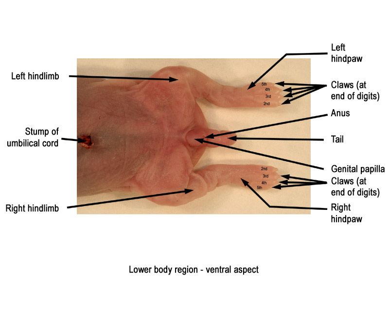
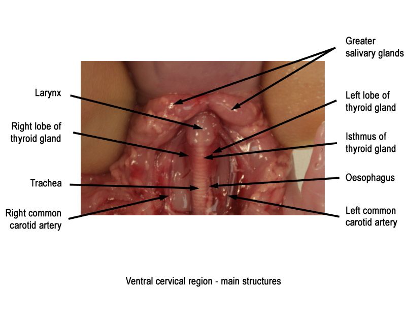
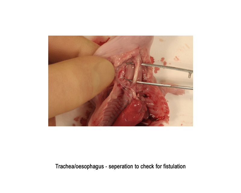
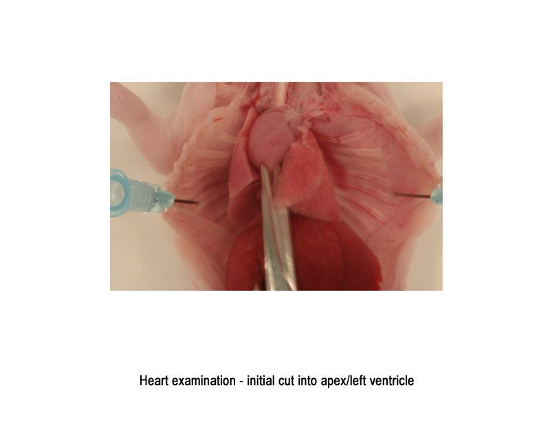
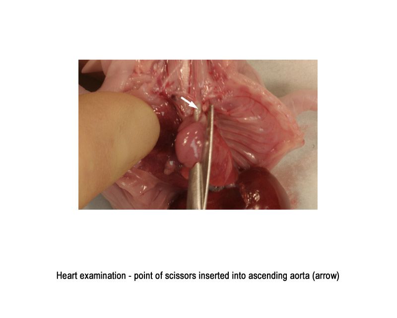
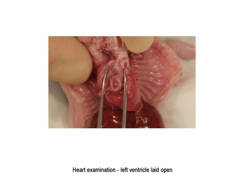
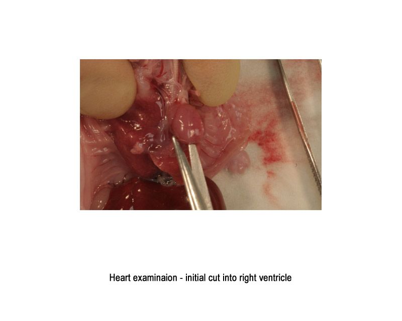
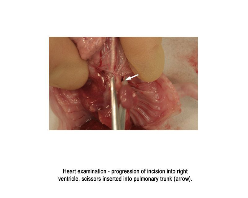
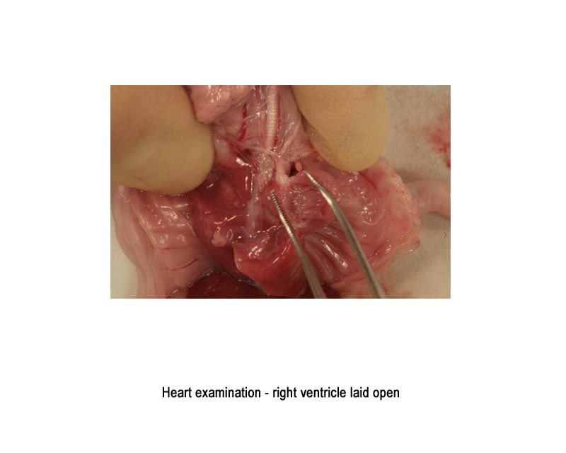
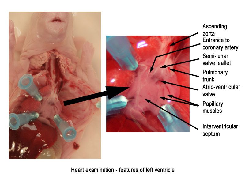
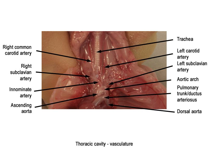
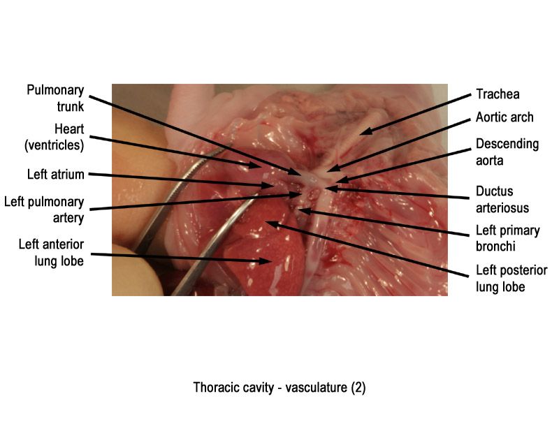
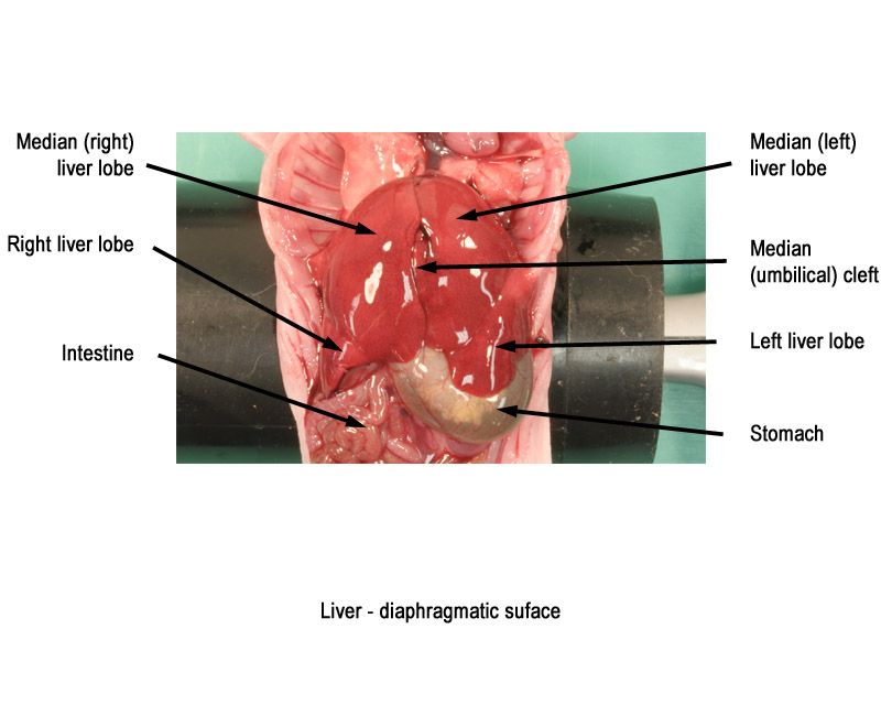
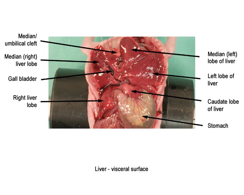
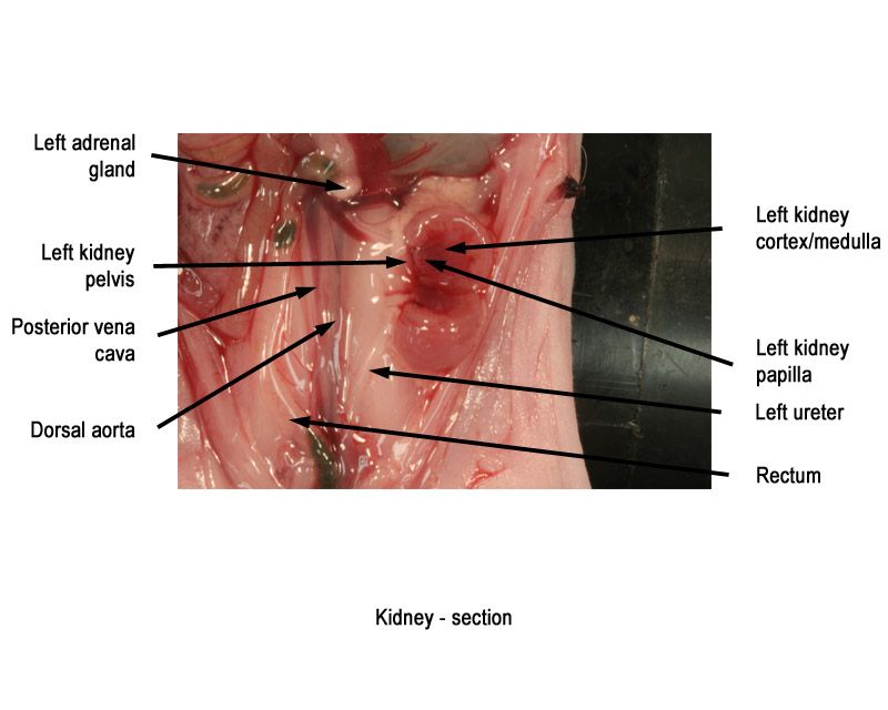
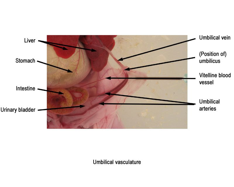
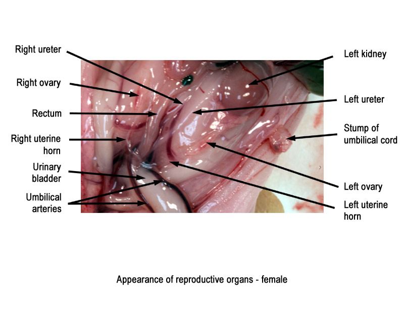
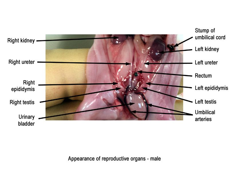
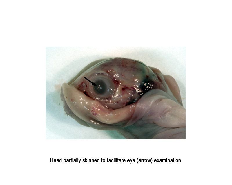
The images below show the normal appearance of the fresh rabbit fetus at Day 29 of gestation (day mating observed = Day 0).
It is recommended that all structures listed on the The International Register of Fetal Morphologists (IRFM) Expected Minimum Structure List for Fetal Morphology Examinations document be examined and that the Fresh external and visceral examination - key structures and notes document be used as a reference.
Learning objective: Compare the image with your own specimens and identify all of the structures that have been labelled.
Click on the images below to open the photo gallery
(Note that the large haemorrhage visible in dorsal scapular region has been caused by a subcutaneous injection of sodium pentobarbitone, used to kill the fetus)


Key learning point exercises and questions
Give 3 reasons why fresh examination is an effective method of examining rabbit fetuses externally/viscerally.
Name 3 requirements that should be in place in the lab before examination of fresh tissues can take place.
What protective clothing should you wear when performing examination of fresh tissues?
Where would you find a list of the head and body areas that should be looked at during external/visceral examination
Use your own specimens to find examples, if possible of:
- Cleft palate
- Subcutaneous haemorrhages
- Absent/small thyroid gland
- Any observations relating to the thymus gland
- Any observations relating to the aortic arch/pulmonary trunk/ductus arteriosus.
- Right subclavian artery arising from aortic arch (absent innominate artery)
- Ventricular septal defects
- What severity levels are they?
- Are they relating to the membranous or muscular area of the septum?
- Any observations relating to the lungs
- Diaphragmatic hernia
- Any observations relating to the liver
- Any observations relating to the kidney and ureter
- What severity levels are they?
- Any observations relating to the gonads
List 2 observations that may be associated with fetuses with subcutaneous oedema
List 3 observations, which would help you distinguish the abnormality situs inversus